Morales-Delgado Nicanor, Castro-Robles Beatriz, Ferrán José L, Martinez-de-la-Torre Margaret, Puelles Luis, Díaz Carmen
Department of Medical Sciences, School of Medicine, Regional Centre for Biomedical Research and Institute for Research in Neurological Disabilities, University of Castilla-La Mancha, Calle Almansa, 14, 02006, Albacete, Spain.
Brain Struct Funct. 2014 May;219(3):1083-111. doi: 10.1007/s00429-013-0554-2. Epub 2013 Apr 30.
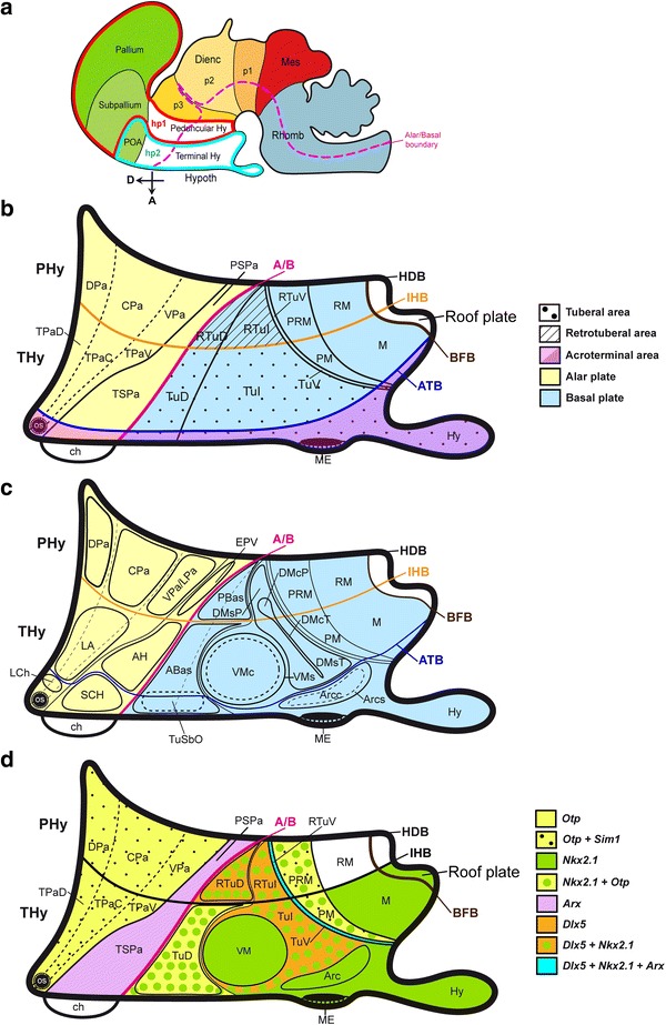
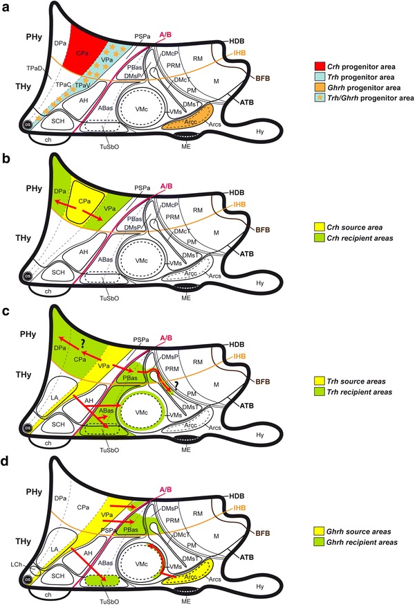
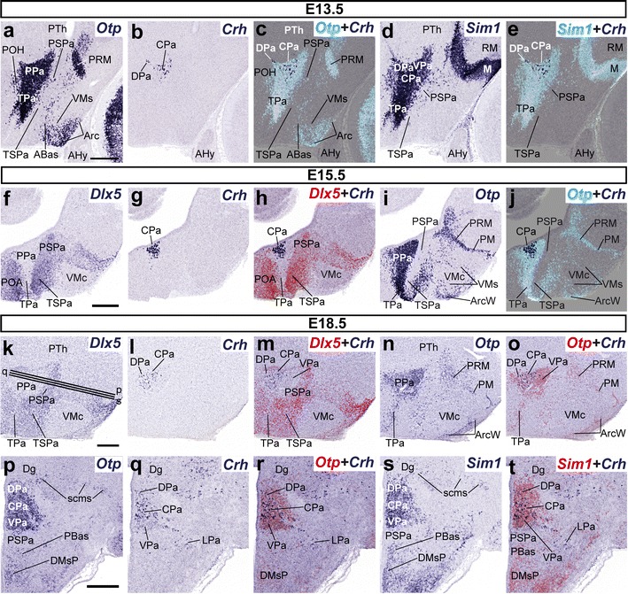
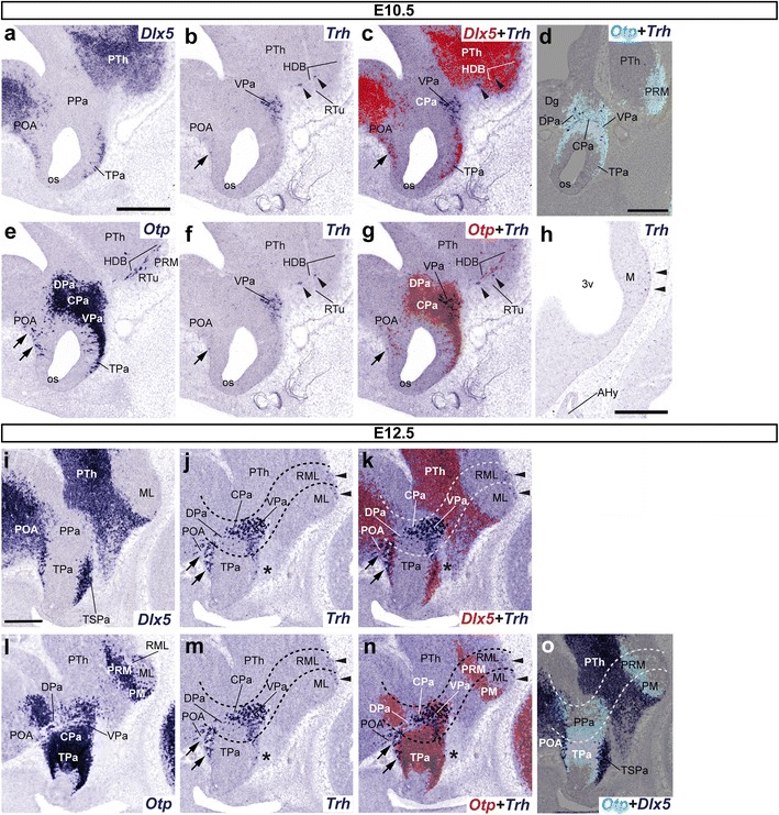
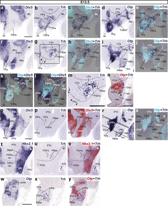
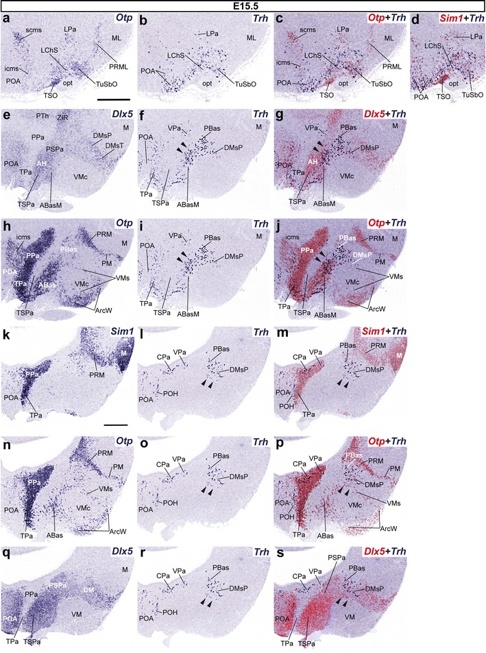
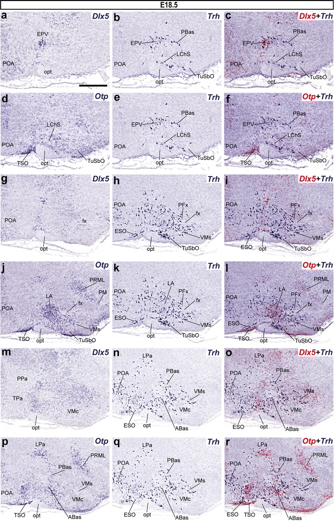
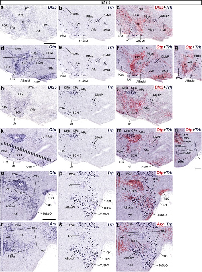
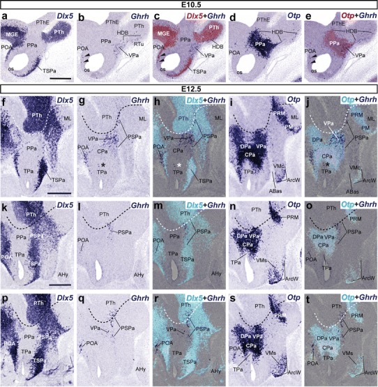
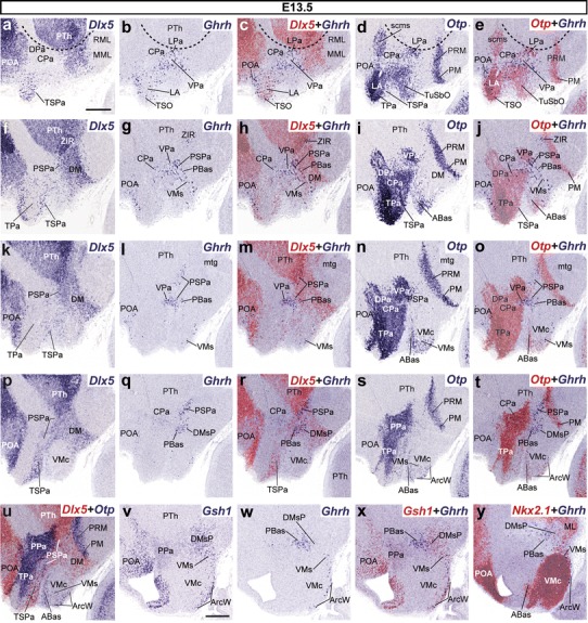
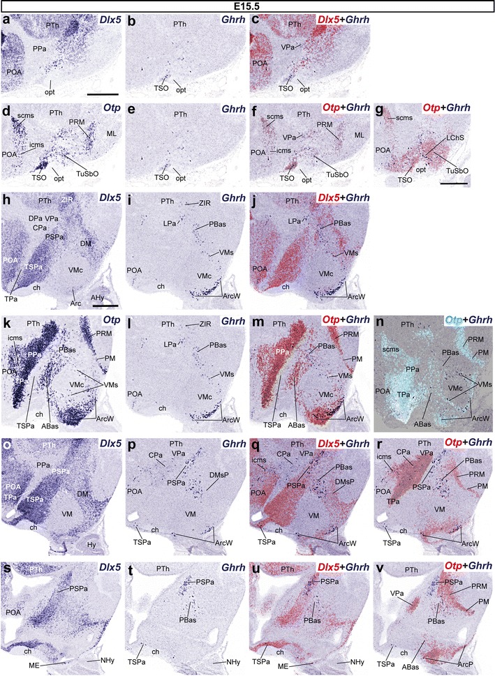
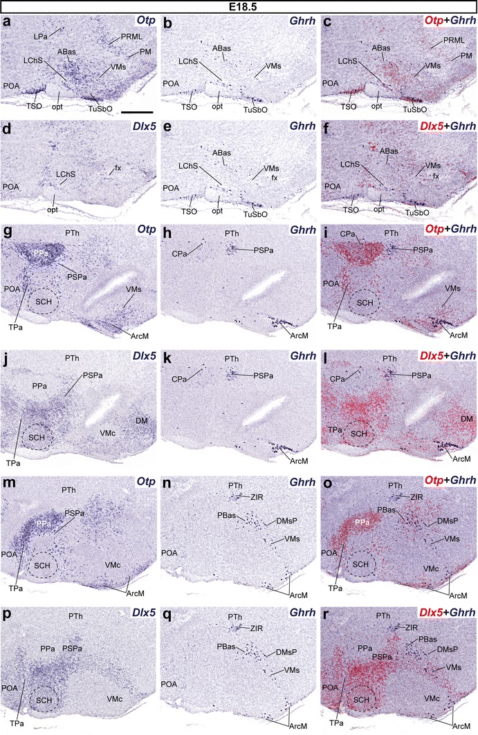
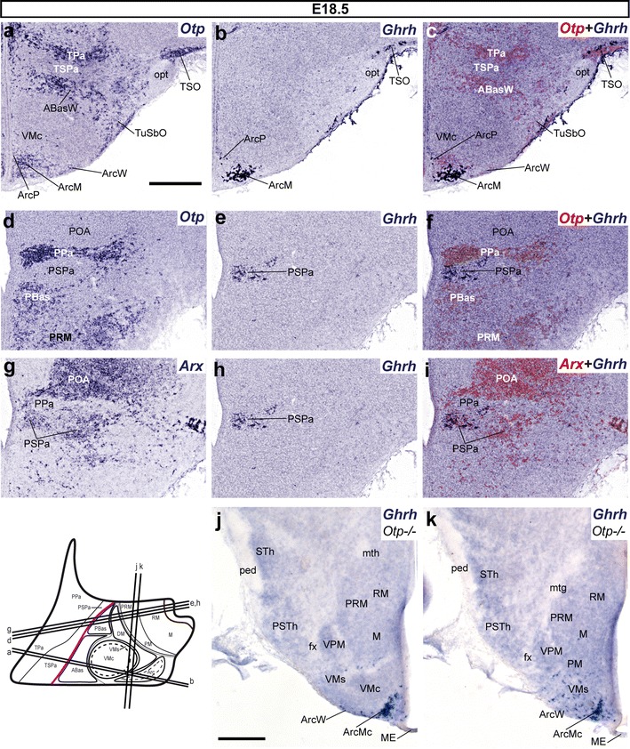
According to the updated prosomeric model, the hypothalamus is subdivided rostrocaudally into terminal and peduncular parts, and dorsoventrally into alar, basal, and floor longitudinal zones. In this context, we examined the ontogeny of peptidergic cell populations expressing Crh, Trh, and Ghrh mRNAs in the mouse hypothalamus, comparing their distribution relative to the major progenitor domains characterized by molecular markers such as Otp, Sim1, Dlx5, Arx, Gsh1, and Nkx2.1. All three neuronal types originate mainly in the peduncular paraventricular domain and less importantly at the terminal paraventricular domain; both are characteristic alar Otp/Sim1-positive areas. Trh and Ghrh cells appeared specifically at the ventral subdomain of the cited areas after E10.5. Additional Ghrh cells emerged separately at the tuberal arcuate area, characterized by Nkx2.1 expression. Crh-positive cells emerged instead in the central part of the peduncular paraventricular domain at E13.5 and remained there. In contrast, as development progresses (E13.5-E18.5) many alar Ghrh and Trh cells translocate into the alar subparaventricular area, and often also into underlying basal neighborhoods expressing Nkx2.1 and/or Dlx5, such as the tuberal and retrotuberal areas, becoming partly or totally depleted at the original birth sites. Our data correlate a topologic map of molecularly defined hypothalamic progenitor areas with three types of specific neurons, each with restricted spatial origins and differential migratory behavior during prenatal hypothalamic development. The study may be useful for detailed causal analysis of the respective differential specification mechanisms. The postulated migrations also contribute to our understanding of adult hypothalamic complexity.
根据更新后的原节段模型,下丘脑在头尾方向上可分为终末部和脚间部,在背腹方向上可分为翼板、基底部和底板纵区。在此背景下,我们研究了小鼠下丘脑中表达促肾上腺皮质激素释放激素(Crh)、促甲状腺激素释放激素(Trh)和生长激素释放激素(Ghrh)mRNA的肽能细胞群的个体发生,比较了它们相对于以Otp、Sim1、Dlx5、Arx、Gsh1和Nkx2.1等分子标记为特征的主要祖域的分布。所有这三种神经元类型主要起源于脚间室旁区,在终末室旁区的起源较少;这两个区域都是典型的翼板Otp/Sim1阳性区域。Trh和Ghrh细胞在胚胎第10.5天(E10.5)后出现在上述区域的腹侧亚区。另外,Ghrh细胞在以Nkx2.1表达为特征的结节弓状区单独出现。Crh阳性细胞在E13.5时出现在脚间室旁区的中央部分,并一直留在那里。相反,随着发育进程(E13.5 - E18.5),许多翼板Ghrh和Trh细胞迁移到翼板室旁下区,并且常常还迁移到表达Nkx2.1和/或Dlx5的下方基底部区域,如结节区和结节后区,导致其在原始产生部位部分或完全耗尽。我们的数据将分子定义的下丘脑祖区拓扑图与三种特定神经元类型相关联,每种神经元类型在产前下丘脑发育过程中具有受限的空间起源和不同的迁移行为。该研究可能有助于对各自的差异特化机制进行详细的因果分析。所推测的迁移也有助于我们理解成年下丘脑的复杂性。