• 文献检索
  • 文档翻译
  • 深度研究
  • 学术资讯
  • Suppr Zotero 插件Zotero 插件
  • 邀请有礼
  • 套餐&价格
  • 历史记录
应用&插件
Suppr Zotero 插件Zotero 插件浏览器插件Mac 客户端Windows 客户端微信小程序
定价
高级版会员购买积分包购买API积分包
服务
文献检索文档翻译深度研究API 文档MCP 服务
关于我们
关于 Suppr公司介绍联系我们用户协议隐私条款
关注我们

Suppr 超能文献

核心技术专利:CN118964589B侵权必究
粤ICP备2023148730 号-1Suppr @ 2026

文献检索

告别复杂PubMed语法,用中文像聊天一样搜索,搜遍4000万医学文献。AI智能推荐,让科研检索更轻松。

立即免费搜索

文件翻译

保留排版,准确专业,支持PDF/Word/PPT等文件格式,支持 12+语言互译。

免费翻译文档

深度研究

AI帮你快速写综述,25分钟生成高质量综述,智能提取关键信息,辅助科研写作。

立即免费体验

代谢改变:一种用于放射性脑损伤的生物标志物——磁共振波谱研究

Metabolic alterations: a biomarker for radiation-induced normal brain injury-an MR spectroscopy study.

作者信息

Sundgren P C, Nagesh V, Elias A, Tsien C, Junck L, Gomez Hassan D M, Lawrence T S, Chenevert T L, Rogers L, McKeever P, Cao Y

机构信息

Department of Radiology, University of Michigan University Health Systems, Ann Arbor, Michigan 48109, USA.

出版信息

J Magn Reson Imaging. 2009 Feb;29(2):291-7. doi: 10.1002/jmri.21657.

DOI:10.1002/jmri.21657
PMID:19161192
原文链接:https://pmc.ncbi.nlm.nih.gov/articles/PMC2679518/
Abstract

PURPOSE

To assess if interval changes in metabolic status in normal cerebral tissue after radiation therapy (RT) can be detected by 2D CSI (chemical shift imaging) proton spectroscopy.

MATERIALS AND METHODS

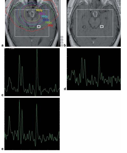
Eleven patients with primary brain tumors undergoing cranial radiation therapy (RT) were included. 2D-CSI MRS was performed before, during, and after the course of RT with the following parameters: TE/TR 144/1500 ms, field of view (FOV) 24, thickness 10 mm, matrix 16 x 16. The metabolic ratios choline/creatine (Cho/Cr), N-acetylaspartate (NAA)/Cr, and NAA/Cho in normal brain tissue were calculated.

RESULTS

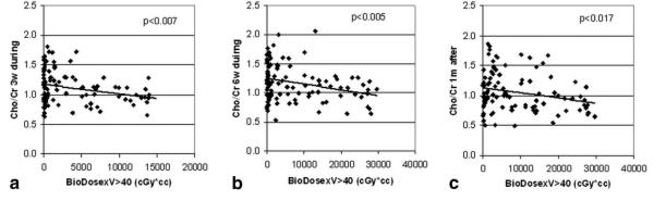
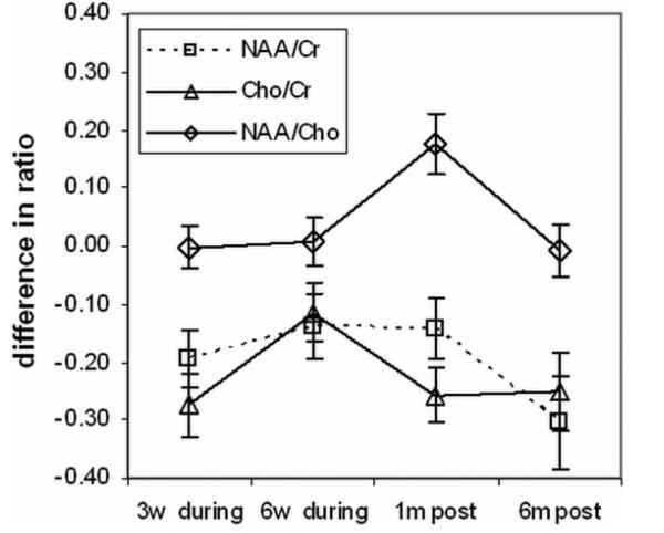
NAA/Cr and Cho/Cr were significantly decreased at week 3 during RT and at 1 month and 6 months after RT compared to values prior to RT (P < 0.01). The NAA/Cr ratio decreased by -0.19 +/- 0.05 (mean +/- standard error [SE]) at week 3 of RT, -0.14 +/- 0.06 at the last week of RT, -0.14 +/- 0.05 at 1 month after RT, and -0.30 +/- 0.08 at 6 months after RT compared to the pre-RT value of 1.43 +/- 0.04. The Cho/Cr ratio decreased by -0.27 +/- 0.05 at week 3 of RT, -0.11 +/- 0.05 at the last week of RT, -0.26 +/- 0.05 at 1 month after RT and -0.25 +/- 0.07 at 6 months after RT from the pre-RT value of 1.29 +/- 0.03. Changes in Cho/Cr were correlated with the interaction of the radiation dose and dose-volume at week 3 of RT, during the last week of RT (P < 0.005), and at 1 month after RT (P = 0.017).

CONCLUSION

The results of this study suggest that MRS can detect early metabolic changes in normal irradiated brain tissue.

摘要

目的

评估二维化学位移成像(2D CSI)质子波谱能否检测放疗(RT)后正常脑组织代谢状态的间期变化。

材料与方法

纳入11例接受颅脑放疗(RT)的原发性脑肿瘤患者。在放疗疗程前、疗程中及疗程后进行二维化学位移成像磁共振波谱(2D-CSI MRS)检查,参数如下:回波时间(TE)/重复时间(TR)为144/1500毫秒,视野(FOV)为24,层厚10毫米,矩阵为16×16。计算正常脑组织中的代谢比值,即胆碱/肌酸(Cho/Cr)、N-乙酰天门冬氨酸(NAA)/Cr和NAA/Cho。

结果

与放疗前相比,放疗第3周、放疗后1个月和6个月时,NAA/Cr和Cho/Cr显著降低(P<0.01)。与放疗前1.43±0.04的值相比,放疗第3周时NAA/Cr比值下降了-0.19±0.05(平均值±标准误差[SE]),放疗最后一周时下降了-0.14±0.06,放疗后1个月时下降了-0.14±0.05,放疗后6个月时下降了-0.30±0.08。与放疗前1.29±0.03的值相比,放疗第3周时Cho/Cr比值下降了-0.27±0.05,放疗最后一周时下降了-0.11±0.05,放疗后1个月时下降了-0.26±0.05,放疗后6个月时下降了-0.25±0.07。在放疗第3周、放疗最后一周(P<0.005)和放疗后1个月(P=0.017)时,Cho/Cr的变化与辐射剂量和剂量体积的相互作用相关。

结论

本研究结果表明,磁共振波谱能够检测正常放疗脑组织的早期代谢变化。

https://cdn.ncbi.nlm.nih.gov/pmc/blobs/6ccd/2679518/be6000884206/nihms-110198-f0003.jpg
https://cdn.ncbi.nlm.nih.gov/pmc/blobs/6ccd/2679518/618f1c505635/nihms-110198-f0001.jpg
https://cdn.ncbi.nlm.nih.gov/pmc/blobs/6ccd/2679518/ccca89977b38/nihms-110198-f0002.jpg
https://cdn.ncbi.nlm.nih.gov/pmc/blobs/6ccd/2679518/be6000884206/nihms-110198-f0003.jpg
https://cdn.ncbi.nlm.nih.gov/pmc/blobs/6ccd/2679518/618f1c505635/nihms-110198-f0001.jpg
https://cdn.ncbi.nlm.nih.gov/pmc/blobs/6ccd/2679518/ccca89977b38/nihms-110198-f0002.jpg
https://cdn.ncbi.nlm.nih.gov/pmc/blobs/6ccd/2679518/be6000884206/nihms-110198-f0003.jpg

相似文献

1
Metabolic alterations: a biomarker for radiation-induced normal brain injury-an MR spectroscopy study.代谢改变:一种用于放射性脑损伤的生物标志物——磁共振波谱研究
J Magn Reson Imaging. 2009 Feb;29(2):291-7. doi: 10.1002/jmri.21657.
2
Transient metabolic changes observed with proton MR spectroscopy in normal human brain after radiation therapy.放疗后正常人类大脑中质子磁共振波谱观察到的短暂代谢变化。
Int J Radiat Oncol Biol Phys. 1998 Jan 15;40(2):279-86. doi: 10.1016/s0360-3016(97)00714-1.
3
MR spectroscopy using normalized and non-normalized metabolite ratios for differentiating recurrent brain tumor from radiation injury.利用归一化和非归一化代谢物比值的磁共振波谱分析鉴别脑肿瘤复发与放射性损伤。
Acad Radiol. 2011 Sep;18(9):1101-8. doi: 10.1016/j.acra.2011.05.006.
4
Differentiation between brain tumor recurrence and radiation injury using MR spectroscopy.利用磁共振波谱区分脑肿瘤复发与放射性损伤。
AJR Am J Roentgenol. 2005 Dec;185(6):1471-6. doi: 10.2214/AJR.04.0933.
5
Multivoxel 3D proton MR spectroscopy in the distinction of recurrent glioma from radiation injury.多体素3D质子磁共振波谱在鉴别复发性胶质瘤与放射性损伤中的应用
J Neurooncol. 2007 Aug;84(1):63-9. doi: 10.1007/s11060-007-9341-3. Epub 2007 Feb 14.
6
Distinction between recurrent glioma and radiation injury using magnetic resonance spectroscopy in combination with diffusion-weighted imaging.利用磁共振波谱结合扩散加权成像鉴别复发性胶质瘤与放射性损伤
Int J Radiat Oncol Biol Phys. 2007 May 1;68(1):151-8. doi: 10.1016/j.ijrobp.2006.12.001. Epub 2007 Feb 7.
7
Long-term Brain Tissue Monitoring after Semi-brain Irradiation in Rats Using Proton Magnetic Resonance Spectroscopy: A Preliminary Study .使用质子磁共振波谱对大鼠半脑照射后的长期脑组织监测:一项初步研究
Chin Med J (Engl). 2017 Apr 20;130(8):957-963. doi: 10.4103/0366-6999.204097.
8
Proton magnetic resonance spectroscopy in the distinction of high-grade cerebral gliomas from single metastatic brain tumors.质子磁共振波谱在鉴别高级别脑胶质瘤与单发性脑转移瘤中的应用
Acta Radiol. 2010 Apr;51(3):316-25. doi: 10.3109/02841850903482901.
9
Proton MR spectroscopy of delayed cerebral radiation in monkeys and humans after brachytherapy.近距离放射治疗后猴子和人类迟发性脑辐射的质子磁共振波谱分析。
AJNR Am J Neuroradiol. 1997 Oct;18(9):1753-61.
10
3-Dimensional magnetic resonance spectroscopic imaging at 3 Tesla for early response assessment of glioblastoma patients during external beam radiation therapy.3特斯拉三维磁共振波谱成像用于胶质母细胞瘤患者外照射放疗期间的早期反应评估
Int J Radiat Oncol Biol Phys. 2014 Sep 1;90(1):181-9. doi: 10.1016/j.ijrobp.2014.05.014. Epub 2014 Jun 28.

引用本文的文献

1
Preclinical monitoring of radiation-induced brain injury via GluCEST MRI and resting-state fMRI at 7 T: an exploratory study on MRI-guided OAR avoidance.通过7T场强下的GluCEST磁共振成像和静息态功能磁共振成像对放射性脑损伤进行临床前监测:一项关于磁共振成像引导下避免危及器官的探索性研究
Strahlenther Onkol. 2025 Apr;201(4):411-419. doi: 10.1007/s00066-024-02292-w. Epub 2024 Sep 11.
2
Brain diffusion MRI biomarkers after oncology treatments.肿瘤治疗后的脑扩散磁共振成像生物标志物
Rep Pract Oncol Radiother. 2024 Feb 16;28(6):823-834. doi: 10.5603/rpor.98728. eCollection 2023.
3
Oxygen supplementation in anesthesia can block FLASH effect and anti-tumor immunity in conventional proton therapy.

本文引用的文献

1
Radiation-induced changes in normal-appearing white matter in patients with cerebral tumors: a diffusion tensor imaging study.脑肿瘤患者正常外观白质的辐射诱导变化:一项扩散张量成像研究。
Int J Radiat Oncol Biol Phys. 2008 Mar 15;70(4):1002-10. doi: 10.1016/j.ijrobp.2007.08.020.
2
Cancer statistics, 2008.2008年癌症统计数据。
CA Cancer J Clin. 2008 Mar-Apr;58(2):71-96. doi: 10.3322/CA.2007.0010. Epub 2008 Feb 20.
3
Quantitative magnetic resonance spectroscopy reveals a potential relationship between radiation-induced changes in rat brain metabolites and cognitive impairment.
麻醉过程中的氧气补充会阻碍传统质子治疗中的FLASH效应和抗肿瘤免疫。
Commun Med (Lond). 2023 Dec 15;3(1):183. doi: 10.1038/s43856-023-00411-9.
4
Delayed effects of radiation in adipose tissue reflect progenitor damage and not cellular senescence.脂肪组织中辐射的延迟效应反映了祖细胞损伤而不是细胞衰老。
Geroscience. 2023 Feb;45(1):507-521. doi: 10.1007/s11357-022-00660-x. Epub 2022 Sep 22.
5
A systematic review on the use of quantitative imaging to detect cancer therapy adverse effects in normal-appearing brain tissue.系统评价使用定量成像技术检测正常外观脑组织中癌症治疗不良反应的研究。
MAGMA. 2022 Feb;35(1):163-186. doi: 10.1007/s10334-021-00985-2. Epub 2021 Dec 17.
6
Early Detection of Radiation-Induced Injury and Prediction of Cognitive Deficit by MRS Metabolites in Radiotherapy of Low-Grade Glioma.磁共振波谱代谢物早期检测及预测低级别胶质瘤放疗后认知功能障碍
Biomed Res Int. 2021 Mar 4;2021:6616992. doi: 10.1155/2021/6616992. eCollection 2021.
7
MR Image Changes of Normal-Appearing Brain Tissue after Radiotherapy.放疗后正常脑组织的磁共振成像变化
Cancers (Basel). 2021 Mar 29;13(7):1573. doi: 10.3390/cancers13071573.
8
Magnetic resonance spectroscopic imaging in gliomas: clinical diagnosis and radiotherapy planning.胶质瘤的磁共振波谱成像:临床诊断与放射治疗计划
BJR Open. 2020 Apr 6;2(1):20190026. doi: 10.1259/bjro.20190026. eCollection 2020.
9
Biomarkers of Brain Damage Induced by Radiotherapy.放疗所致脑损伤的生物标志物
Dose Response. 2020 Jul 9;18(3):1559325820938279. doi: 10.1177/1559325820938279. eCollection 2020 Jul-Sep.
10
Radiation Induced Metabolic Alterations Associate With Tumor Aggressiveness and Poor Outcome in Glioblastoma.辐射诱导的代谢改变与胶质母细胞瘤的肿瘤侵袭性和不良预后相关。
Front Oncol. 2020 May 5;10:535. doi: 10.3389/fonc.2020.00535. eCollection 2020.
定量磁共振波谱分析揭示了大鼠脑代谢物辐射诱导变化与认知障碍之间的潜在关系。
Radiat Res. 2007 Nov;168(5):574-81. doi: 10.1667/RR0735.1.
4
Quantitative in vivo proton MR spectroscopic evaluation of the irradiated rat brain.辐照大鼠脑的定量体内质子磁共振波谱评估
J Magn Reson Imaging. 2007 Dec;26(6):1590-5. doi: 10.1002/jmri.21095.
5
Prognostic significance of serial magnetic resonance spectroscopies over the course of radiation therapy for patients with malignant glioma.恶性胶质瘤患者放射治疗过程中连续磁共振波谱的预后意义。
Clin Invest Med. 2006 Oct;29(5):301-11.
6
Delayed neurotoxicity associated with therapy for children with acute lymphoblastic leukemia.与急性淋巴细胞白血病患儿治疗相关的迟发性神经毒性。
Ment Retard Dev Disabil Res Rev. 2006;12(3):174-83. doi: 10.1002/mrdd.20113.
7
White matter anisotropy in post-treatment childhood cancer survivors: preliminary evidence of association with neurocognitive function.儿童癌症幸存者治疗后的白质各向异性:与神经认知功能关联的初步证据。
J Clin Oncol. 2006 Feb 20;24(6):884-90. doi: 10.1200/JCO.2005.02.4505.
8
Differentiation between brain tumor recurrence and radiation injury using MR spectroscopy.利用磁共振波谱区分脑肿瘤复发与放射性损伤。
AJR Am J Roentgenol. 2005 Dec;185(6):1471-6. doi: 10.2214/AJR.04.0933.
9
Radiation-induced brain metabolic changes in the acute and early delayed phase detected with quantitative proton magnetic resonance spectroscopy.利用定量质子磁共振波谱检测辐射诱导的急性和早期延迟期脑代谢变化。
J Comput Assist Tomogr. 2005 May-Jun;29(3):293-7. doi: 10.1097/01.rct.0000161422.95625.8a.
10
Neuropsychological evaluation of late-onset post-radiotherapy encephalopathy: a comparison with vascular dementia.迟发性放疗后脑病的神经心理学评估:与血管性痴呆的比较
J Neurol Sci. 2005 Mar 15;229-230:195-200. doi: 10.1016/j.jns.2004.11.027. Epub 2004 Dec 19.