Chang Ji-Min, Yoo Young Sam, Kim Dong-Won
Department of Thoracic and Cardiovascular Surgery, Inje University Sanggye Paik Hospital, Korea.
Korean J Thorac Cardiovasc Surg. 2011 Oct;44(5):368-72. doi: 10.5090/kjtcs.2011.44.5.368. Epub 2011 Oct 6.
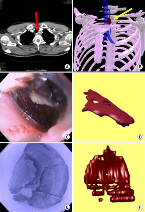
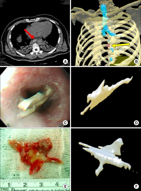
This study was conducted to investigate the clinical application of three-dimensional (3D) reconstructed computed tomography (CT) images in detecting and gaining information on esophageal foreign bodies (FBs). Two patients with esophageal FBs were enrolled for analysis. In both cases, 3D reconstructed images were compared with the FB that was removed according to the object shape, size, location, and orientation in the esophagus. The results indicate the usefulness of conversion of CT data to 3D images to help in diagnosis and treatment. Use of 3D images prior to treatment allows for rapid prototyping and surgery simulation.
本研究旨在探讨三维(3D)重建计算机断层扫描(CT)图像在检测食管异物(FBs)及获取相关信息方面的临床应用。纳入两名食管异物患者进行分析。在这两个病例中,将3D重建图像与根据食管内异物的形状、大小、位置和方向取出的异物进行比较。结果表明,将CT数据转换为3D图像有助于诊断和治疗。治疗前使用3D图像可实现快速成型和手术模拟。