Liu Jia, Xu Jie, Zhou Jun, Zhang Yu, Guo Dajing, Wang Zhigang
Department of Radiology.
Department of Ultrasound, Institute of Ultrasound Imaging, The Second Affiliated Hospital of Chongqing Medical University, Yuzhong, Chongqing, People's Republic of China.
Int J Nanomedicine. 2017 Feb 9;12:1113-1126. doi: 10.2147/IJN.S123228. eCollection 2017.
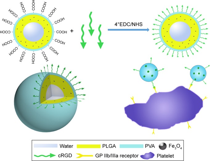
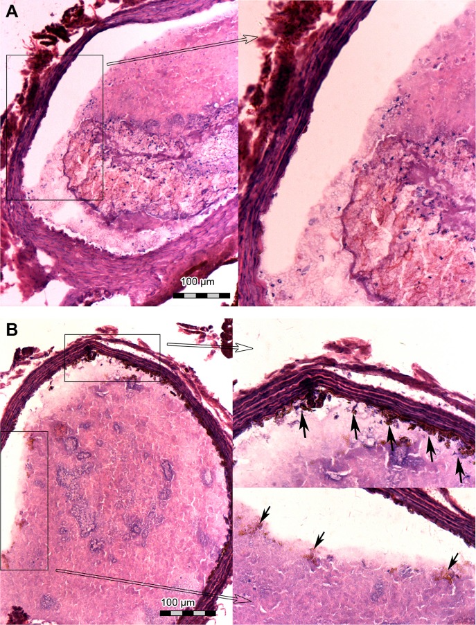
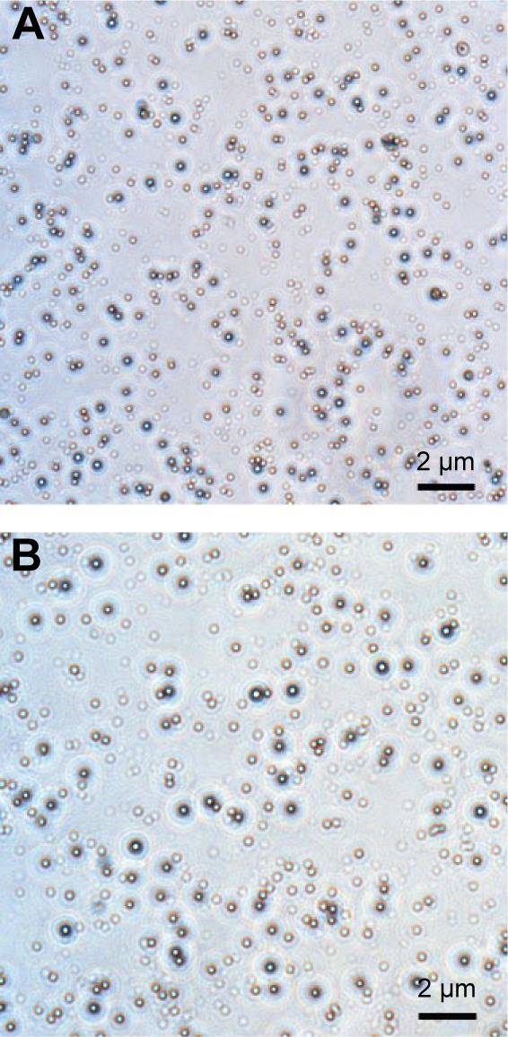
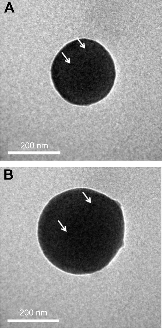
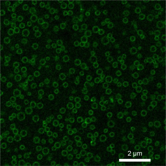
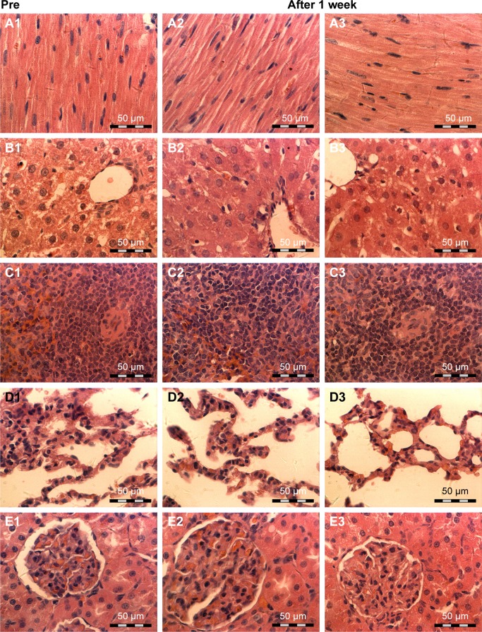
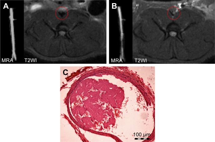
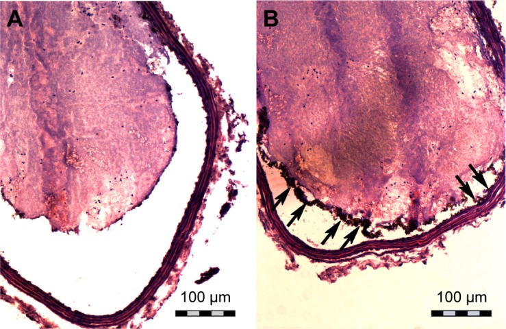
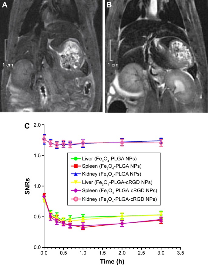
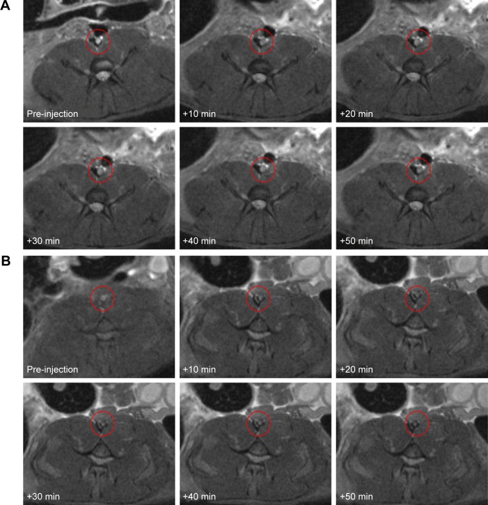
Thrombotic disease is a great threat to human health, and early detection is particularly important. Magnetic resonance (MR) molecular imaging provides noninvasive imaging with the potential for early disease diagnosis. In this study, we developed FeO-based poly(lactic--glycolic acid) (PLGA) nanoparticles (NPs) surface-modified with a cyclic Arg-Gly-Asp (cRGD) peptide as an MR contrast agent for the detection of thrombosis. The physical and chemical characteristics, biological toxicity, ability to target thrombi, and biodistribution of the NPs were studied. The FeO-PLGA-cRGD NPs were constructed successfully, and hematologic and pathologic assays indicated no in vivo toxicity of the NPs. In a rat model of FeCl-induced abdominal aorta thrombosis, the NPs readily and selectively accumulated on the surface of the thrombosis and under vascular endothelial cells ex vivo and in vivo. In the in vivo experiment, the biodistribution of the NPs suggested that the NPs might be internalized by the macrophages of the reticuloendothelial system in the liver and the spleen. The T2 signal decreased at the mural thrombus 10 min after injection and then gradually increased until 50 min. These results suggest that the NPs are suitable for in vivo molecular imaging of thrombosis under high shear stress conditions and represent a very promising MR contrast agent for sensitive and specific detection of thrombosis.
血栓性疾病对人类健康构成巨大威胁,早期检测尤为重要。磁共振(MR)分子成像提供了具有早期疾病诊断潜力的非侵入性成像。在本研究中,我们开发了用环Arg-Gly-Asp(cRGD)肽表面修饰的基于FeO的聚乳酸-乙醇酸共聚物(PLGA)纳米颗粒(NPs)作为用于检测血栓形成的MR造影剂。研究了NPs的物理和化学特性、生物毒性、靶向血栓的能力以及生物分布。成功构建了FeO-PLGA-cRGD NPs,血液学和病理学检测表明NPs在体内无毒性。在FeCl诱导的大鼠腹主动脉血栓形成模型中,NPs在体外和体内均能迅速且选择性地聚集在血栓表面和血管内皮细胞下方。在体内实验中,NPs的生物分布表明NPs可能被肝脏和脾脏中网状内皮系统的巨噬细胞内化。注射后10分钟,壁血栓处的T2信号降低,然后逐渐升高直至50分钟。这些结果表明,NPs适用于在高剪切应力条件下对血栓形成进行体内分子成像,是一种非常有前景的用于敏感和特异性检测血栓形成的MR造影剂。