Holmes Holly E, Powell Nick M, Ma Da, Ismail Ozama, Harrison Ian F, Wells Jack A, Colgan Niall, O'Callaghan James M, Johnson Ross A, Murray Tracey K, Ahmed Zeshan, Heggenes Morten, Fisher Alice, Cardoso M Jorge, Modat Marc, O'Neill Michael J, Collins Emily C, Fisher Elizabeth M C, Ourselin Sébastien, Lythgoe Mark F
Division of Medicine, UCL Centre for Advanced Biomedical Imaging, University College LondonLondon, UK.
Centre for Medical Image Computing, University College LondonLondon, UK.
Front Neuroinform. 2017 Mar 31;11:20. doi: 10.3389/fninf.2017.00020. eCollection 2017.
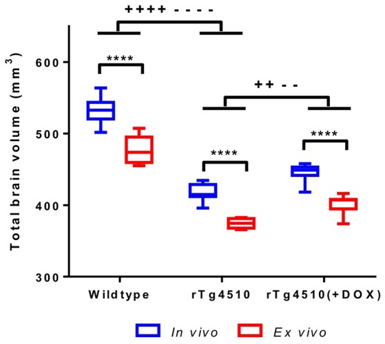
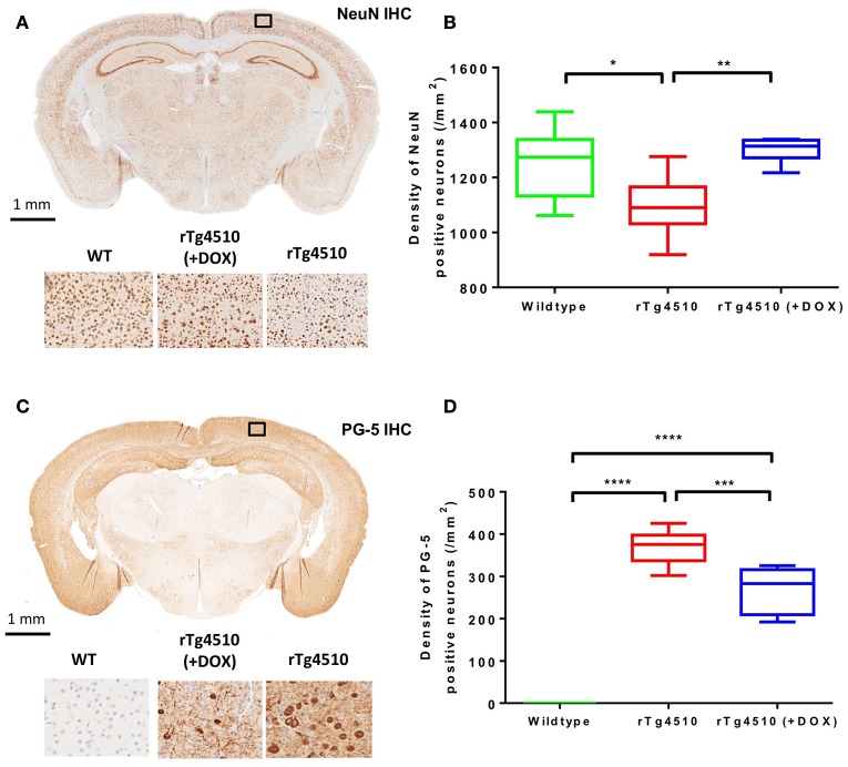
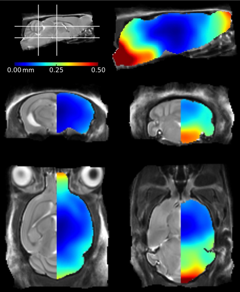
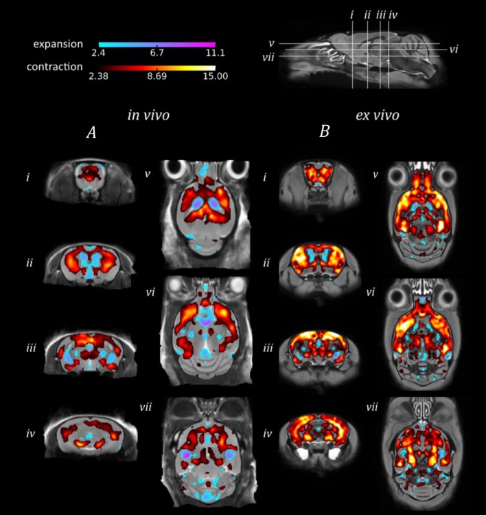
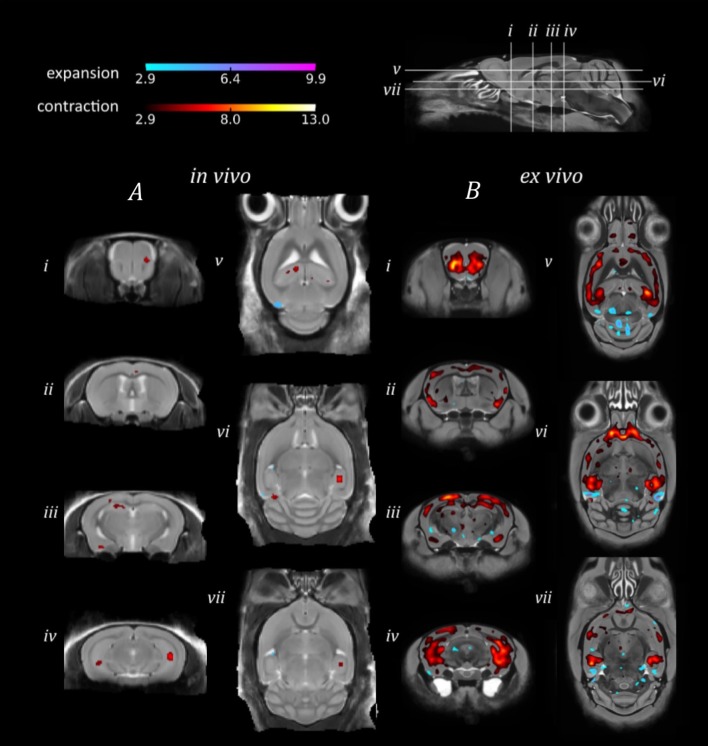
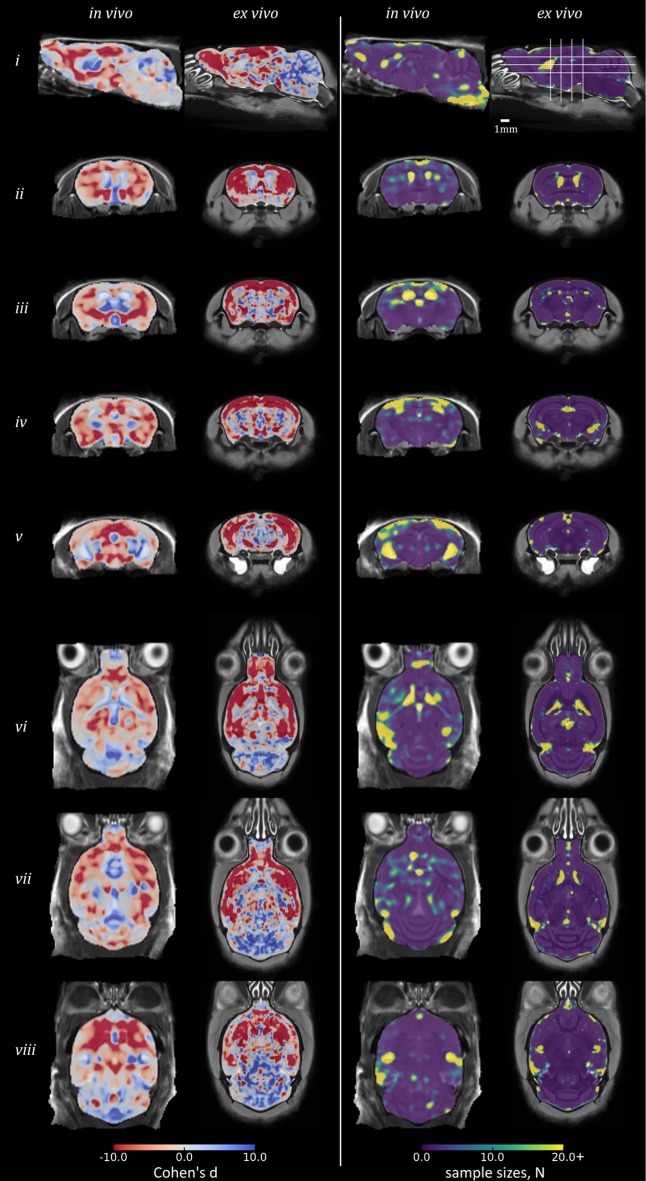
With increasingly large numbers of mouse models of human disease dedicated to MRI studies, compromises between and MRI must be fully understood in order to inform the choice of imaging methodology. We investigate the application of high resolution and MRI, in combination with tensor-based morphometry (TBM), to uncover morphological differences in the rTg4510 mouse model of tauopathy. The rTg4510 mouse also offers a novel paradigm by which the overexpression of mutant tau can be regulated by the administration of doxycycline, providing us with a platform on which to investigate more subtle alterations in morphology with morphometry. Both and MRI allowed the detection of widespread bilateral patterns of atrophy in the rTg4510 mouse brain relative to wild-type controls. Regions of volume loss aligned with neuronal loss and pathological tau accumulation demonstrated by immunohistochemistry. When we sought to investigate more subtle structural alterations in the rTg4510 mice relative to a subset of doxycycline-treated rTg4510 mice, imaging enabled the detection of more regions of morphological brain changes. The disadvantages of MRI may however mitigate this increase in sensitivity: we observed a 10% global shrinkage in brain volume of the post-mortem tissues due to formalin fixation, which was most notable in the cerebellum and olfactory bulbs. However, many central brain regions were not adversely affected by the fixation protocol, perhaps due to our "in-skull" preparation. The disparity between our TBM findings from and MRI underlines the importance of appropriate study design, given the trade-off between these two imaging approaches. We support the utility of MRI for morphological phenotyping of mouse models of disease; however, for subtler phenotypes, offers enhanced sensitivity to discrete morphological changes.
随着越来越多致力于磁共振成像(MRI)研究的人类疾病小鼠模型的出现,为了指导成像方法的选择,必须充分了解不同类型MRI之间的权衡。我们研究了高分辨率结构和功能MRI与基于张量的形态测量(TBM)相结合的应用,以揭示tau蛋白病的rTg4510小鼠模型中的形态学差异。rTg4510小鼠还提供了一种新的范例,通过给予强力霉素可以调节突变tau蛋白的过表达,为我们提供了一个用形态测量法研究更细微形态学改变的平台。与野生型对照相比,结构和功能MRI均能检测到rTg4510小鼠脑内广泛的双侧萎缩模式。体积缩小区域与免疫组织化学显示的神经元丢失和病理性tau蛋白积累一致。当我们试图研究rTg4510小鼠相对于一部分接受强力霉素治疗的rTg4510小鼠更细微的结构改变时,功能成像能够检测到更多脑形态学变化区域。然而,结构MRI的缺点可能会减轻这种敏感性的增加:我们观察到死后组织的脑体积因福尔马林固定而整体缩小了10%,这在小脑和嗅球中最为明显。然而,许多脑中央区域并未受到固定方案的不利影响,这可能是由于我们的“颅骨内”制备方法。鉴于这两种成像方法之间的权衡,我们在结构和功能MRI上的TBM结果差异凸显了适当研究设计的重要性。我们支持结构MRI在疾病小鼠模型形态学表型分析中的应用;然而,对于更细微的表型,功能成像对离散形态学变化具有更高的敏感性。