Emory Vaccine Center, Emory University, Atlanta, GA, United States.
Department of Microbiology and Immunology, Yerkes National Primate Research Center, Emory University, Atlanta, GA, United States.
Front Immunol. 2020 Jan 17;10:3053. doi: 10.3389/fimmu.2019.03053. eCollection 2019.
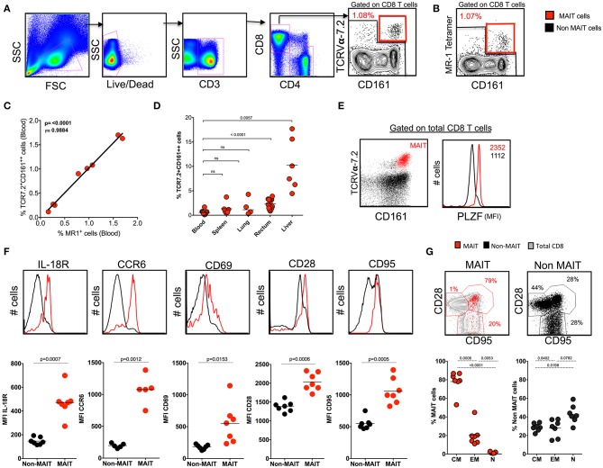
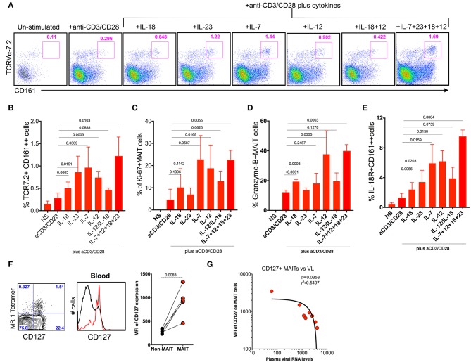
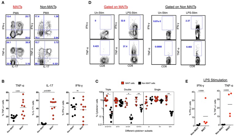
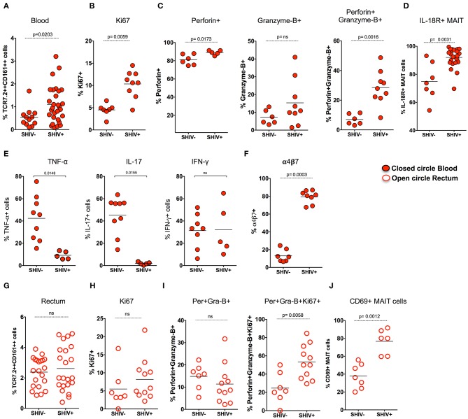
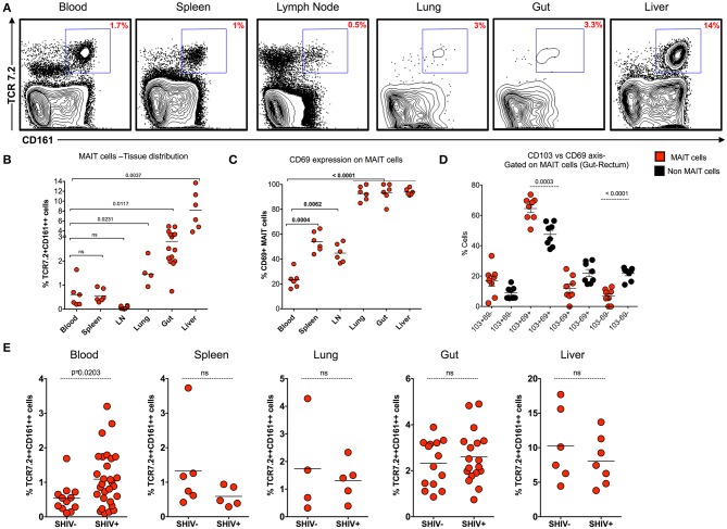
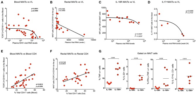
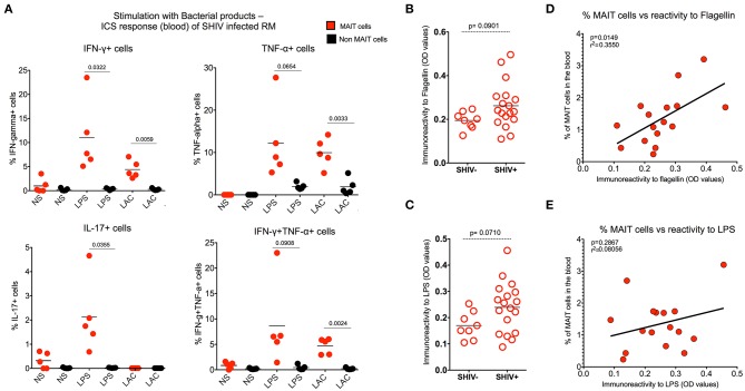
Mucosa-associated invariant T (MAIT) cells are recently characterized as a novel subset of innate-like T cells that recognize microbial metabolites as presented by the MHC-1b-related protein MR1. The significance of MAIT cells in anti-bacterial defense is well-understood but not clear in viral infections such as SIV/HIV infection. Here we studied the phenotype, distribution, and function of MAIT cells and their association with plasma viral levels during chronic SHIV infection in rhesus macaques (RM). Two groups of healthy and chronic SHIV-infected macaques were characterized for MAIT cells in blood and mucosal tissues. Similar to human, we found a significant fraction of macaque T cells co-expressing MAIT cell markers CD161 and TCRVα-7.2 that correlated directly with macaque MR1 tetramer. These cells displayed memory phenotype and expressed high levels of IL-18R, CCR6, CD28, and CD95. During chronic infection, the frequency of MAIT cells are enriched in the blood but unaltered in the rectum; both blood and rectal MAIT cells displayed higher proliferative and cytotoxic phenotype post-SHIV infection. The frequency of MAIT cells in blood and rectum correlated inversely with plasma viral RNA levels and correlated directly with total CD4 T cells. MAIT cells respond to microbial products during chronic SHIV infection and correlated positively with serum immunoreactivity to flagellin levels. Tissue distribution analysis of MAIT cells during chronic infection showed significant enrichment in the non-lymphoid tissues (lung, rectum, and liver) compared to lymphoid tissues (spleen and LN), with higher levels of tissue-resident markers CD69 and CD103. Exogenous cytokine treatments during chronic SHIV infection revealed that IL-7 is important for the proliferation of MAIT cells, but IL-12 and IL-18 are important for their cytolytic function. Overall our results demonstrated that MAIT cells are enriched in blood but unaltered in the rectum during chronic SHIV infection, which displayed proliferative and functional phenotype that inversely correlated with SHIV plasma viral RNA levels. Treatment such as combined cytokine treatments could be beneficial for enhancing functional MAIT cells during chronic HIV infection .
黏膜相关不变 T(MAIT)细胞最近被鉴定为一种新型的先天样 T 细胞亚群,能够识别 MHC-1b 相关蛋白 MR1 呈递的微生物代谢物。MAIT 细胞在抗细菌防御中的作用已经得到很好的理解,但在 SIV/HIV 感染等病毒感染中尚不清楚。在这里,我们研究了 MAIT 细胞的表型、分布和功能,以及它们与慢性 SIV 感染恒河猴(RM)血浆病毒水平的关联。我们对两组健康和慢性 SIV 感染的猕猴进行了 MAIT 细胞的特征分析,包括血液和黏膜组织。与人类相似,我们发现猕猴 T 细胞中有相当一部分表达 MAIT 细胞标志物 CD161 和 TCRVα-7.2,这些细胞与猕猴 MR1 四聚体直接相关。这些细胞表现出记忆表型,表达高水平的 IL-18R、CCR6、CD28 和 CD95。在慢性感染期间,MAIT 细胞在血液中的频率增加,但在直肠中没有改变;血液和直肠 MAIT 细胞在感染 SIV 后表现出更高的增殖和细胞毒性表型。血液和直肠 MAIT 细胞的频率与血浆病毒 RNA 水平呈负相关,与总 CD4 T 细胞呈正相关。MAIT 细胞在慢性 SIV 感染期间对微生物产物有反应,并与血清对鞭毛蛋白水平的免疫反应呈正相关。慢性感染期间 MAIT 细胞的组织分布分析显示,与淋巴组织(脾和淋巴结)相比,非淋巴组织(肺、直肠和肝脏)中 MAIT 细胞明显富集,组织驻留标志物 CD69 和 CD103 水平较高。在慢性 SIV 感染期间进行的外源性细胞因子治疗显示,IL-7 对 MAIT 细胞的增殖很重要,而 IL-12 和 IL-18 对其细胞毒性功能很重要。总的来说,我们的结果表明,MAIT 细胞在慢性 SIV 感染期间在血液中富集,但在直肠中不变,其增殖和功能表型与 SIV 血浆病毒 RNA 水平呈负相关。联合细胞因子治疗等治疗方法可能有助于在慢性 HIV 感染期间增强功能性 MAIT 细胞。