Department of Biomaterials, Institute of Clinical Sciences, Sahlgrenska Academy, University of Gothenburg, Gothenburg, Sweden.
The Brånemark Clinic, Public Dental Service, Region Västra Götaland, Gothenburg, Sweden.
Clin Exp Dent Res. 2021 Apr;7(2):137-146. doi: 10.1002/cre2.344. Epub 2020 Nov 9.
This clinical randomized study aimed to evaluate the early plaque formation on nonresorbable polytetrafluoroethylene (PTFE) membranes having either a dense (d-PTFE) or an expanded (e-PTFE) microstructure and exposed to the oral cavity.
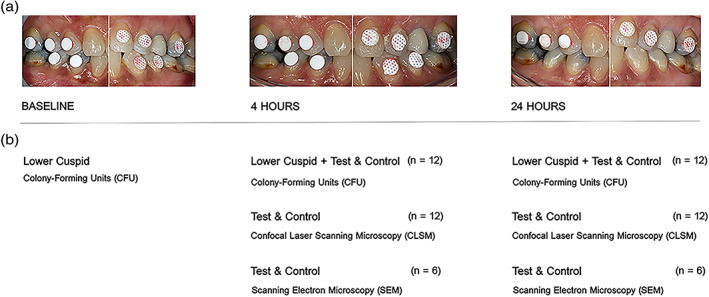
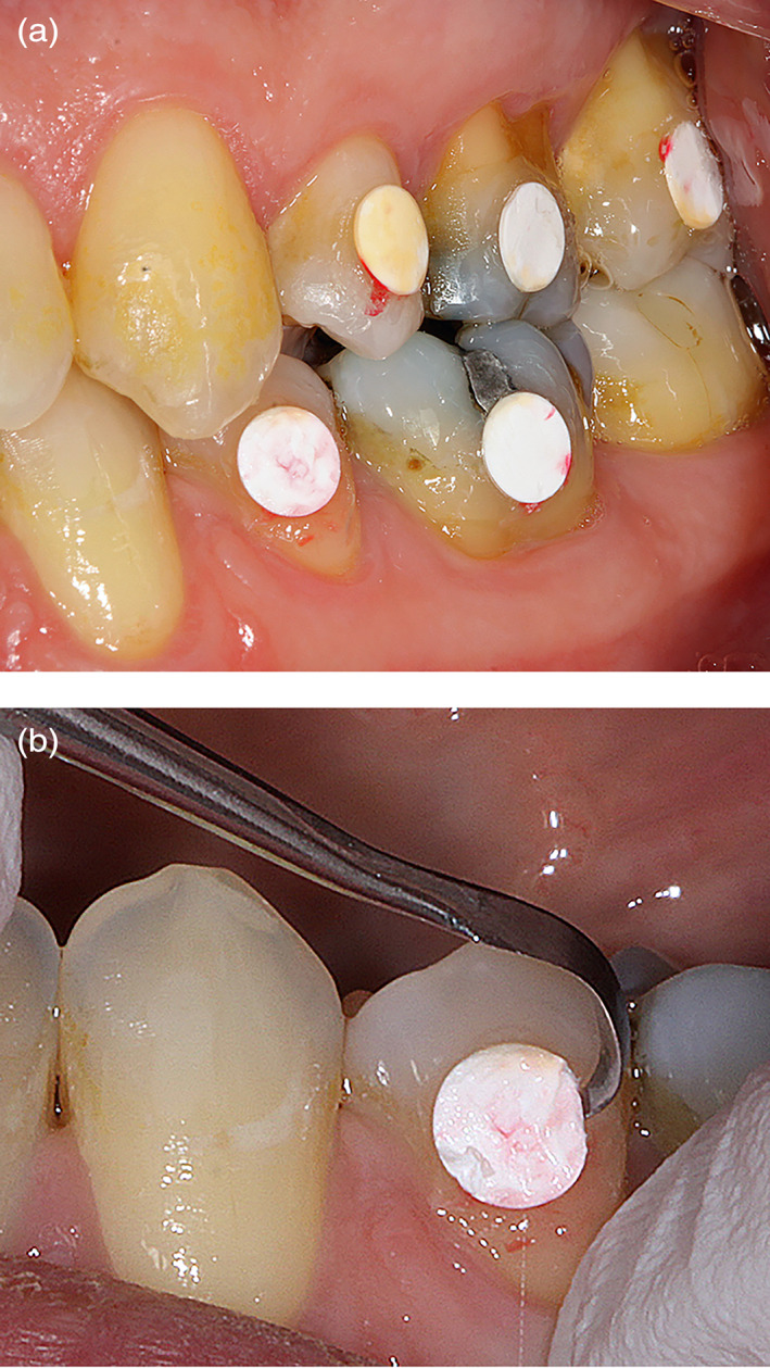
Twelve individuals were enrolled in this study. In a split-mouth design, five test membranes (e-PTFE) with a dual-layer configuration and five control membranes (d-PTFE) were bonded on the buccal surfaces of posterior teeth of each subject. All study subjects refrained from toothbrushing during the study period. Specimens were detached from the teeth at 4 and 24 hr and subjected to viability counting, confocal microscopy, and scanning electron microscopy. Plaque samples were harvested from neighboring teeth at baseline, 4, and 24 hr, as control. Wilcoxon signed rank test was applied.
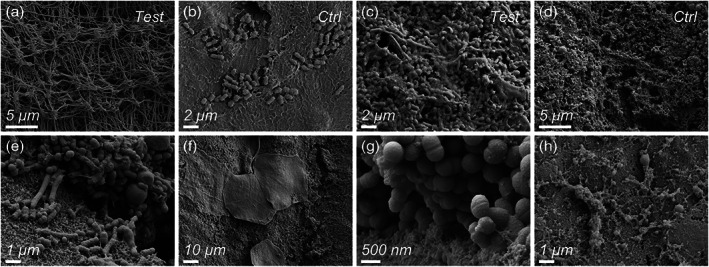
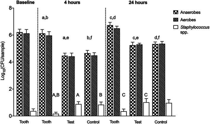
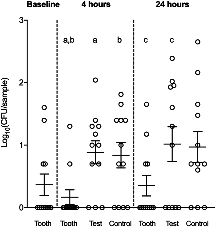
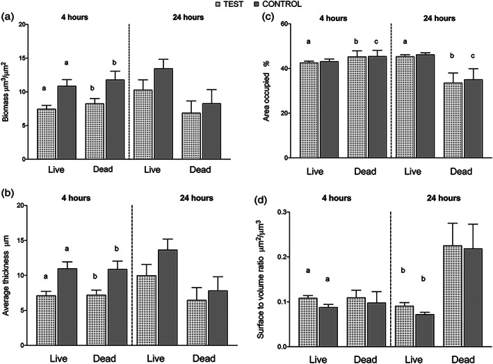
No bond failure of the membranes was reported. Between the early and late time points, viable bacterial counts increased on all membranes, with no difference between the test and control. The number of Staphylococcus spp. decreased on the tooth surfaces and increased on both membranes overtime, with a significant difference compared to teeth. The total biomass and average biofilm thickness of live and dead cells were significantly greater at the d-PTFE barriers after 4 hr.
This study demonstrated that the e-PTFE membrane was associated with a lesser degree of biofilm accumulation during the initial exposure compared to the d-PTFE membrane. The present experimental setup provides a valuable toolbox to study the in vivo behavior of different membranes used in guided bone regeneration (GBR).
本临床随机研究旨在评估暴露于口腔环境中的不可吸收聚四氟乙烯(PTFE)致密(d-PTFE)或膨体(e-PTFE)微观结构膜的早期菌斑形成情况。
本研究纳入了 12 名个体。采用分口设计,将 5 个测试膜(e-PTFE)的双层结构和 5 个对照膜(d-PTFE)粘结在每个受试者的后牙颊面。所有研究受试者在研究期间均避免刷牙。在 4 小时和 24 小时时从牙齿上取下标本进行活菌计数、共聚焦显微镜和扫描电子显微镜检查。在基线、4 小时和 24 小时时,从相邻牙齿上采集菌斑样本作为对照。应用 Wilcoxon 符号秩检验。
未报告膜的粘结失败。在早期和晚期时间点之间,所有膜上的活菌计数均增加,但测试膜与对照膜之间无差异。在牙齿表面,葡萄球菌属的数量减少,而在两种膜上随时间增加,与牙齿相比有显著差异。4 小时后,d-PTFE 屏障上的活细胞和死细胞的总生物量和平均生物膜厚度显著更高。
本研究表明,与 d-PTFE 膜相比,e-PTFE 膜在初始暴露期间与较少的生物膜积聚相关。本实验设置为研究不同用于引导骨再生(GBR)的膜的体内行为提供了一个有价值的工具包。