Department of Neurosurgery, Eberhard Karls University Tübingen, Hoppe-Seyler-Strasse 3, 72076, Tübingen, Germany.
Department of Neuroradiology, Eberhard Karls University Tübingen, Tübingen, Germany.
Neurosurg Rev. 2021 Dec;44(6):3459-3469. doi: 10.1007/s10143-021-01521-5. Epub 2021 Mar 22.
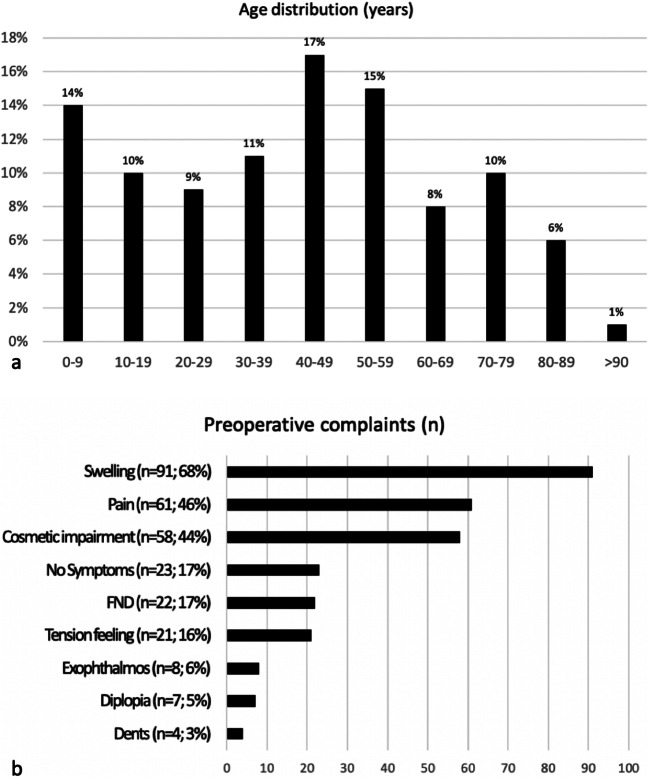
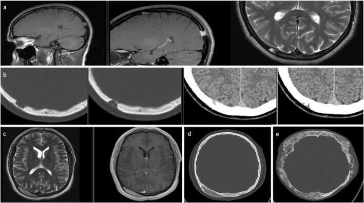
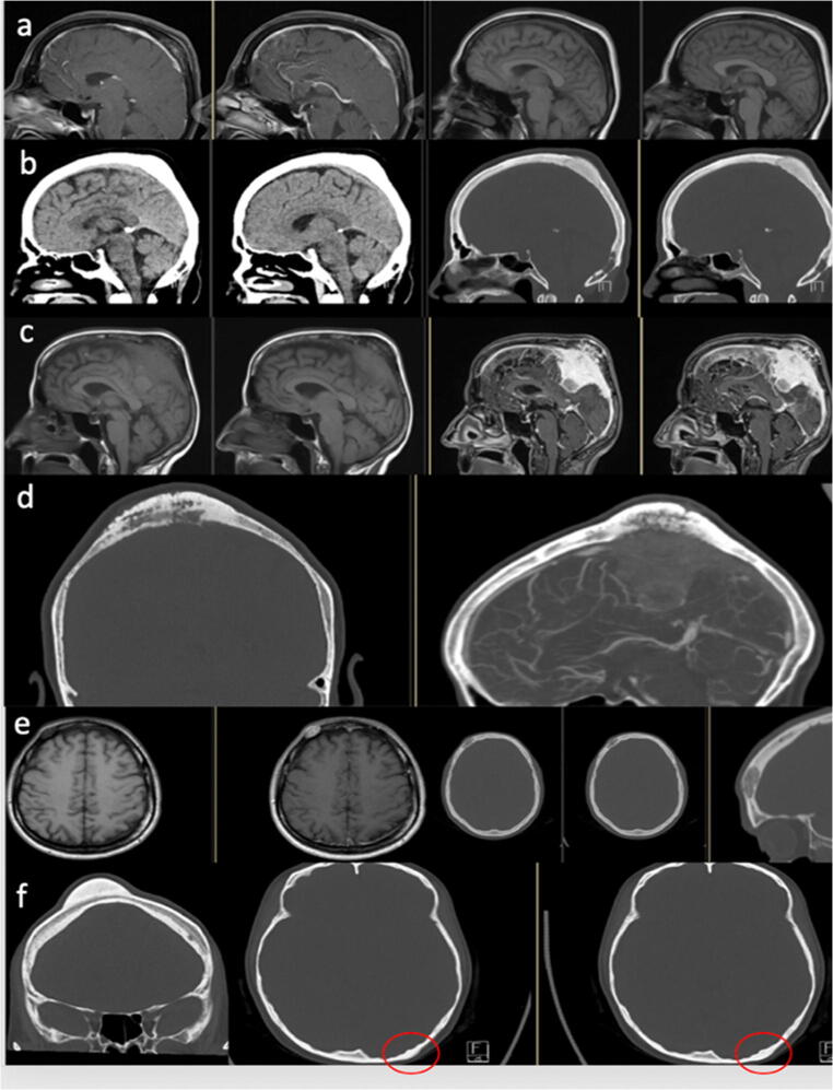
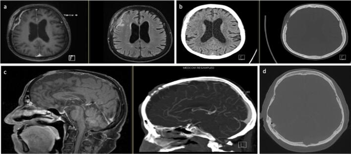
Calvarial lesions are rare and can present as a variety of different diseases. The lesions can be palpable on the skin and cause local pain and paraesthesia and, depending on the location, neurological deficits can also occur. This research aims to present an overview of typical imaging features as well as neurosurgical management. We examined the charts of patients who underwent surgery on a calvarial lesion in our department between 2004 and 2017 (n=133). Retrospectively, the pre-, intra-, and postoperative data were analyzed with morphological and histological findings and compared with each other. Pain, swelling, cosmetically disturbing, and neurological deficits were the main complaints. Seventy-seven lesions were limited to the bone, while another 56 lesions showed an infiltrating growth in the adjacent tissue. Depending on the clinical signs and suspected diagnosis, a biopsy, a partial removal, or a complete resection was performed. Histiocytosis (n=20), meningiomas (n=20), metastases (n=19), and osteomas (n=16) were the most common lesions. Fibrous dysplasia (n=6) and intraosseous hemangioma (n=9) were less common; other lesions were present only in isolated cases. Imaging features may suggest the lesion to be benign or malignant, but the diagnosis can be only confirmed by histological examination. The surgical strategy depends on the complaints, location of the lesion, and suspected diagnosis. Adjuvant treatment should be initiated according to the histological findings.
颅骨病变较为罕见,可表现为多种不同疾病。病变可在皮肤表面触及,引起局部疼痛和感觉异常,且根据病变位置的不同,还可能出现神经功能缺损。本研究旨在介绍颅骨病变的典型影像学特征和神经外科治疗方法。我们回顾性分析了 2004 年至 2017 年期间在我科接受颅骨病变手术的患者的病历(n=133)。对术前、术中及术后的资料进行形态学和组织学检查,并与其他结果进行对比。主要症状包括疼痛、肿胀、美容性困扰和神经功能缺损。77 例病变局限于骨,另有 56 例病变向邻近组织浸润性生长。根据临床表现和疑似诊断,进行活检、部分切除或完全切除。组织学检查显示,病变以组织细胞增生症(n=20)、脑膜瘤(n=20)、转移瘤(n=19)和骨瘤(n=16)最为常见,纤维结构不良(n=6)和骨内血管瘤(n=9)较为少见,其他病变则较为罕见。影像学特征有助于提示病变的良恶性,但最终诊断仍需依靠组织学检查。手术策略取决于患者的症状、病变位置和疑似诊断。应根据组织学检查结果选择辅助治疗方案。