The International Peace Maternity and Child Health Hospital, School of Medicine, Shanghai Jiao Tong University, 1961 Hua-Shan Road, Shanghai, 200030, People's Republic of China.
School of Biomedical Engineering, Shanghai Jiao Tong University, 1954 Hua-Shan Road, Shanghai, 200030, People's Republic of China.
Stem Cell Res Ther. 2021 Apr 1;12(1):223. doi: 10.1186/s13287-021-02280-2.
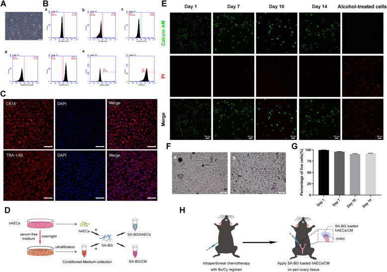
Human amniotic epithelial cells (hAECs) exhibit a strong capability to restore ovarian function in chemotherapy-induced premature ovarian failure (POF). However, the therapeutic efficacy of hAECs is usually affected by the limited number and proliferative ability of grafted hAECs in target organs. The transplantation of stem cells encapsulated in sodium alginate-bioglass (SA-BG) composite hydrogel has recently been shown to be an effective strategy for tissue regeneration. The current study aims to investigate the therapeutic potential of hAECs or hAEC-derived conditioned medium (CM) encapsulated in SA-BG in mice with chemotherapy-induced POF.
C57BL/6 mice were intraperitoneally injected with chemotherapy drugs to induce POF. hAECs or CM were harvested and encapsulated in SA-BG composite hydrogel, which were transplanted onto the injured ovaries of mice with POF. Follicle development, granulosa cell function, and ovarian angiogenesis were evaluated by morphological methods. To further elucidate the effect of SA-BG-encapsulated hAECs/CM on vascularization, the tube formation of human umbilical vein epithelial cells (hUVECs) was conducted in vitro. Cytokine array and ELISA were used to analyze and quantify the effects of bioactive components released by SA-BG on the secretion of angiogenic factors by hAECs.
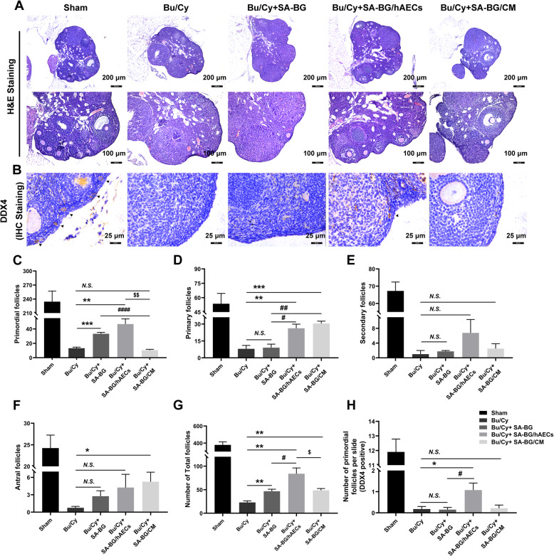
The transplantation of SA-BG-encapsulated hAECs/CM restored follicle development, repaired granulosa cell function, and enhanced ovarian angiogenesis in POF mice. The further study showed that SA-BG significantly promoted the tube formation of hUVECs in vitro. Moreover, encapsulating hAECs could facilitate the effect of SA-BG on inducing the formation of the capillary tube in a paracrine manner. In addition, we found that SA-BG extracts significantly enhanced the viability of hAECs and stimulated the secretion of pro-angiogenic factors of hAECs. Notably, compared with SA-BG/CM, SA-BG/hAECs achieve better therapeutic effects, possibly because stimulation of BG enhanced the viability and paracrine capacity of hAECs.
The present study initially demonstrates that SA-BG-encapsulated hAECs or CM can exert a therapeutic effect on chemotherapy-induced POF mainly by protecting granulosa cell function and enhancing ovarian vascularization, which might provide a novel strategy for the delivery of hAECs for treating POF.
人羊膜上皮细胞(hAECs)在化疗诱导的卵巢早衰(POF)中表现出很强的恢复卵巢功能的能力。然而,移植到靶器官中的 hAECs 的数量和增殖能力有限,通常会影响其治疗效果。最近的研究表明,将干细胞包封在海藻酸钠-生物玻璃(SA-BG)复合水凝胶中进行移植是一种有效的组织再生策略。本研究旨在探讨 hAECs 或 hAEC 来源的条件培养基(CM)包封在 SA-BG 中对化疗诱导的 POF 小鼠的治疗潜力。
C57BL/6 小鼠经腹腔注射化疗药物诱导 POF。收获 hAECs 或 CM 并包封在 SA-BG 复合水凝胶中,移植到 POF 小鼠的受损卵巢上。通过形态学方法评估卵泡发育、颗粒细胞功能和卵巢血管生成。为了进一步阐明 SA-BG 包封的 hAECs/CM 对血管生成的影响,在体外进行了人脐静脉上皮细胞(hUVECs)的管形成实验。采用细胞因子阵列和 ELISA 分析和定量 SA-BG 对 hAECs 分泌血管生成因子的生物活性成分的影响。
SA-BG 包封的 hAECs/CM 移植恢复了 POF 小鼠的卵泡发育,修复了颗粒细胞功能,增强了卵巢血管生成。进一步的研究表明,SA-BG 显著促进了 hUVECs 在体外的管形成。此外,包封 hAECs 可以促进 SA-BG 以旁分泌方式诱导毛细血管形成的作用。此外,我们发现 SA-BG 提取物显著提高了 hAECs 的活力,并刺激了 hAECs 分泌促血管生成因子。值得注意的是,与 SA-BG/CM 相比,SA-BG/hAECs 实现了更好的治疗效果,这可能是因为 BG 的刺激增强了 hAECs 的活力和旁分泌能力。
本研究初步表明,SA-BG 包封的 hAECs 或 CM 可以通过保护颗粒细胞功能和增强卵巢血管生成来发挥对化疗诱导的 POF 的治疗作用,这可能为 hAECs 治疗 POF 提供一种新的递送策略。