Department of Neurosurgery, Johns Hopkins University, Baltimore, MD 21287, United States; Department of Neural and Pain Sciences, University of Maryland School of Dentistry, Baltimore, MD 21201, United States; Center to Advance Chronic Pain Research, University of Maryland Baltimore, Baltimore, MD 21201, United States.
Department of Neurosurgery, Johns Hopkins University, Baltimore, MD 21287, United States; Department of Neural and Pain Sciences, University of Maryland School of Dentistry, Baltimore, MD 21201, United States; Center to Advance Chronic Pain Research, University of Maryland Baltimore, Baltimore, MD 21201, United States; Clinical Neuroengineering, BrainMind Institute and Centre of Neuroprosthetics (CNP), Swiss Federal Institute of Technology (EPFL) Geneva, Switzerland and Swiss Federal Institute of Technology Valais (EPFL Valais), Sion, Switzerland; WyssCenter of Bio- and Neuroengineering, Geneva, Switzerland.
Neuroimage. 2022 Aug 1;256:119278. doi: 10.1016/j.neuroimage.2022.119278. Epub 2022 May 9.
Resting state functional connectivity (FC) is widely used to assess functional brain alterations in patients with chronic pain. However, reports of FC accompanying tonic pain in pain-free persons are rare. A network we term the Descending Pain Modulatory Network (DPMN) is implicated in healthy and pathologic pain modulation. Here, we evaluate the effect of tonic pain on FC of specific nodes of this network: anterior cingulate cortex (ACC), amygdala (AMYG), periaqueductal gray (PAG), and parabrachial nuclei (PBN).
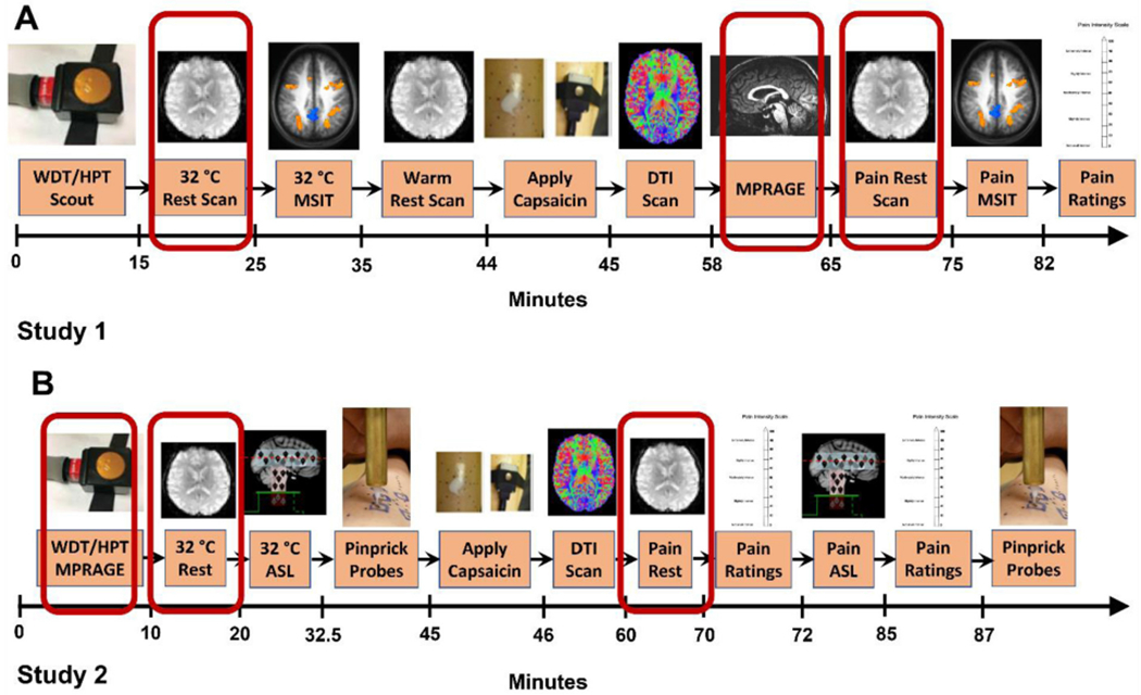
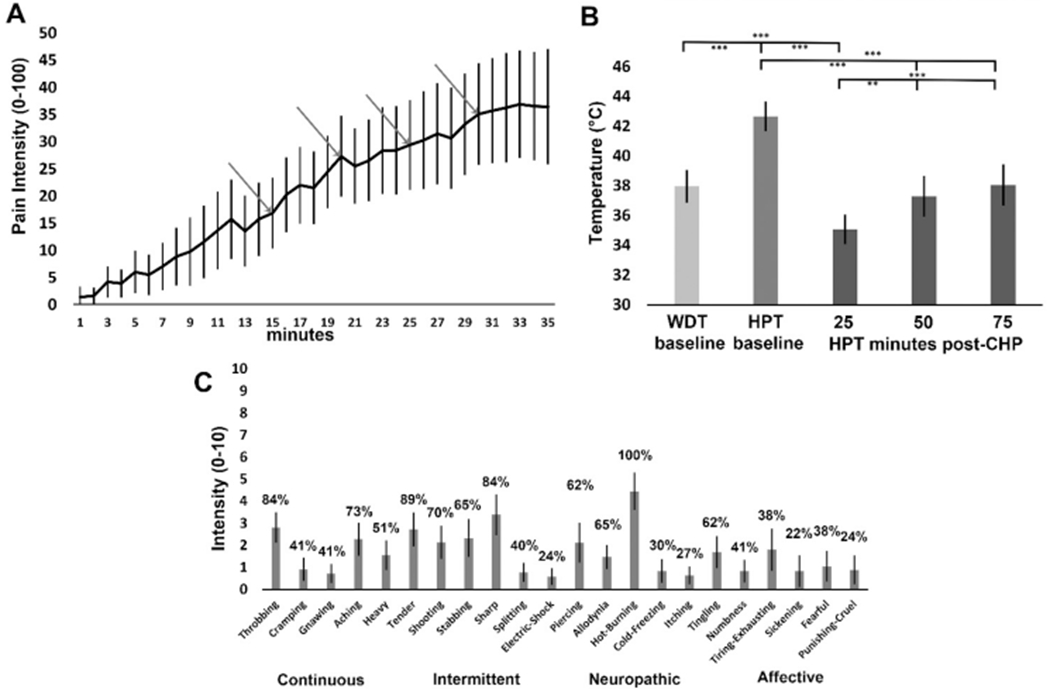
In 50 pain-free participants (30F), we induced tonic pain using a capsaicin-heat pain model. functional MRI measured resting BOLD signal during pain-free rest with a 32 °C thermode and then tonic pain where participants experienced a previously warm temperature combined with capsaicin. We evaluated FC from ACC, AMYG, PAG, and PBN with correlation of self-report pain intensity during both states. We hypothesized tonic pain would diminish FC dyads within the DPMN.
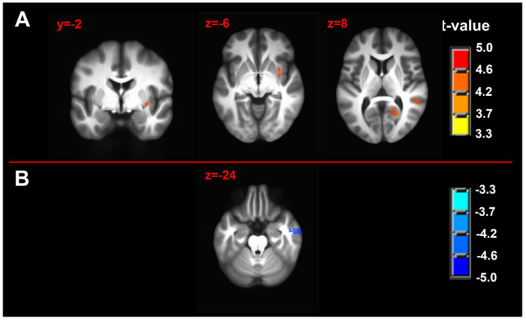
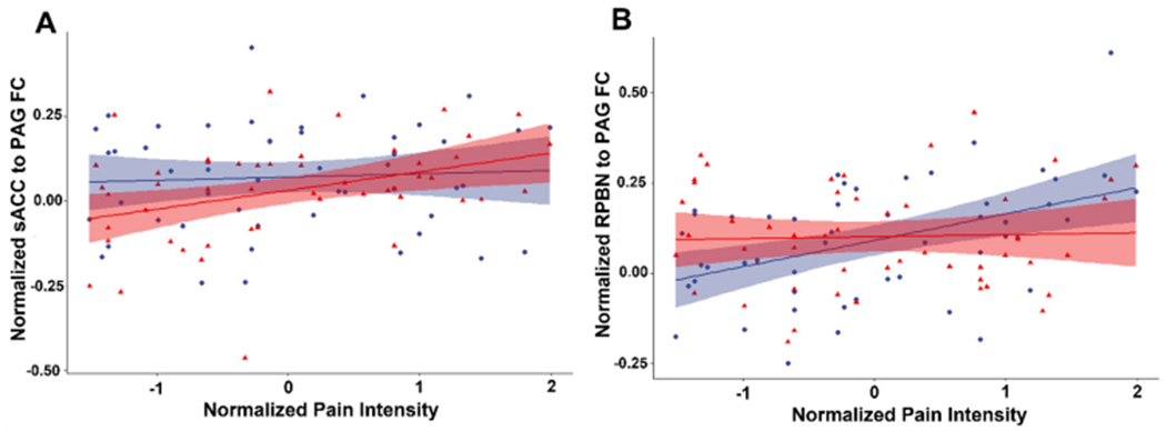
Of all hypothesized FC dyads, only PAG and subgenual ACC was weakly altered during pain (F = 3.34; p = 0.074; pain-free>pain d = 0.25). After pain induction sACC-PAG FC became positively correlated with pain intensity (R = 0.38; t = 2.81; p = 0.007). Right PBN-PAG FC during pain-free rest positively correlated with subsequently experienced pain (R = 0.44; t = 3.43; p = 0.001). During pain, this connection's FC was diminished (paired t=-3.17; p = 0.0026). In whole-brain analyses, during pain-free rest, FC between left AMYG and right superior parietal lobule and caudate nucleus were positively correlated with subsequent pain. During pain, FC between left AMYG and right inferior temporal gyrus negatively correlated with pain. Subsequent pain positively correlated with right AMYG FC with right claustrum; right primary visual cortex and right temporo-occipitoparietal junction CONCLUSION: We demonstrate sACC-PAG tonic pain FC positively correlates with experienced pain and resting right PBN-PAG FC correlates with subsequent pain and is diminished during tonic pain. Finally, we reveal PAG- and right AMYG-anchored networks which correlate with subsequently experienced pain intensity. Our findings suggest specific connectivity patterns within the DPMN at rest are associated with subsequently experienced pain and modulated by tonic pain. These nodes and their functional modulation may reveal new therapeutic targets for neuromodulation or biomarkers to guide interventions.
静息态功能连接(FC)广泛用于评估慢性疼痛患者的大脑功能改变。然而,在无痛人群中报告与紧张性疼痛相关的 FC 却很少见。一个我们称之为下行疼痛调制网络(DPMN)的网络被认为与健康和病理性疼痛调制有关。在这里,我们评估了紧张性疼痛对该网络特定节点的 FC 的影响:前扣带皮层(ACC)、杏仁核(AMYG)、导水管周围灰质(PAG)和臂旁核(PBN)。
在 50 名无痛参与者(30 名女性)中,我们使用辣椒素热痛模型诱导紧张性疼痛。在使用 32°C 热模型无痛休息期间,功能 MRI 测量静息时的 BOLD 信号,然后在参与者经历先前温暖的温度和辣椒素的情况下进行紧张性疼痛。我们用自我报告的两种状态下的疼痛强度相关性来评估 ACC、AMYG、PAG 和 PBN 的 FC。我们假设紧张性疼痛会减少 DPMN 内的 FC 二联体。
在所假设的所有 FC 二联体中,只有 PAG 和 subgenual ACC 在疼痛时发生微弱改变(F=3.34;p=0.074;无痛>疼痛 d=0.25)。在疼痛诱导后,sACC-PAG FC 与疼痛强度呈正相关(R=0.38;t=2.81;p=0.007)。右 PBN-PAG 在无痛休息时的 FC 与随后经历的疼痛呈正相关(R=0.44;t=3.43;p=0.001)。在疼痛期间,这种连接的 FC 减少(配对 t=-3.17;p=0.0026)。在全脑分析中,在无痛休息期间,左杏仁核和右顶叶上回和尾状核之间的 FC 与随后的疼痛呈正相关。在疼痛期间,左杏仁核和右颞下回之间的 FC 与疼痛呈负相关。随后的疼痛与右杏仁核与右屏状核、右初级视皮层和右颞枕顶叶联合区的 FC 呈正相关。
我们证明了 sACC-PAG 紧张性疼痛 FC 与经历的疼痛呈正相关,而右 PBN-PAG 在静息时的 FC 与随后的疼痛相关,并在紧张性疼痛时减少。最后,我们揭示了与随后经历的疼痛强度相关的以 PAG 和右杏仁核为中心的网络。我们的发现表明,DPNM 在休息时的特定连接模式与随后经历的疼痛有关,并受紧张性疼痛的调节。这些节点及其功能调节可能为神经调节或生物标志物提供新的治疗靶点,以指导干预。