Ji Zhao-Jie, Shi Yun, Li Xing, Hou Rui, Yang Yu, Liu Zhu-Qing, Duan Xian-Chun, Liu Qing, Chen Wei-Dong, Peng Dai-Yin
School of Pharmacy, Anhui University of Chinese Medicine, Hefei, China.
Anhui Province Key Laboratory of Chinese Medicinal Formula, Anhui University of Chinese Medicine, Hefei, China.
Front Pharmacol. 2022 Jun 8;13:910217. doi: 10.3389/fphar.2022.910217. eCollection 2022.
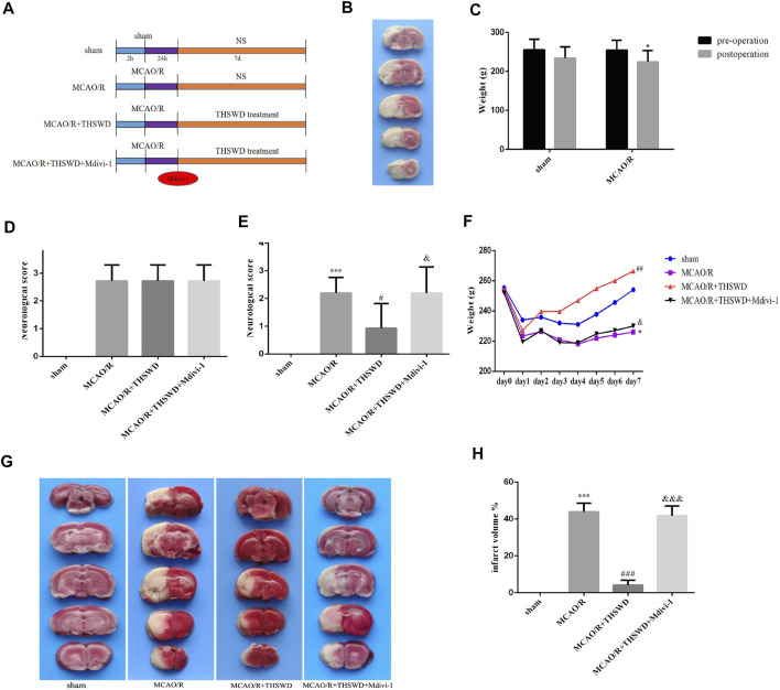
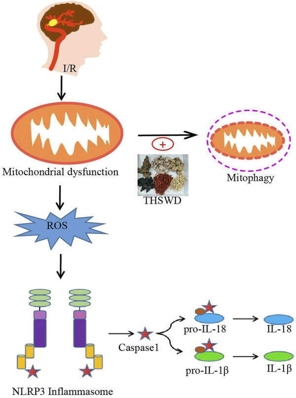
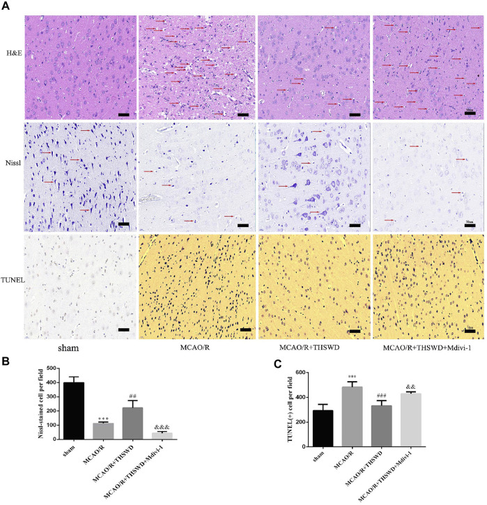
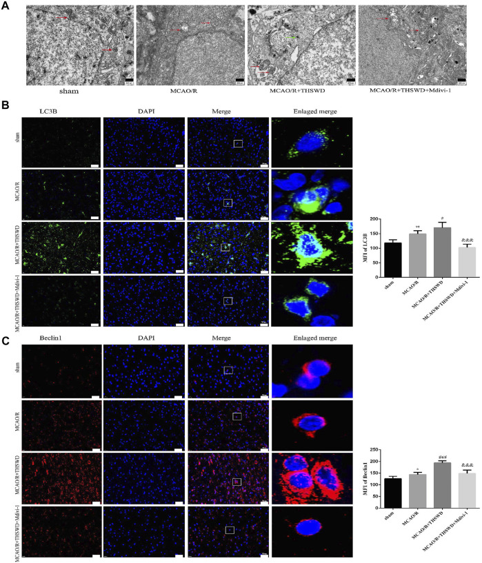
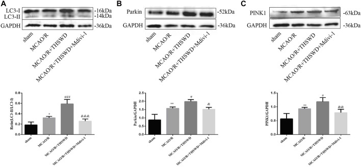
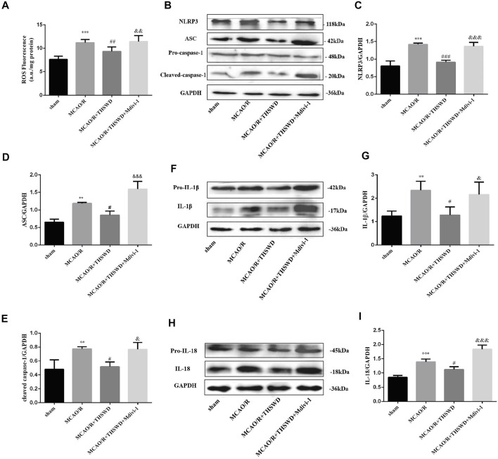
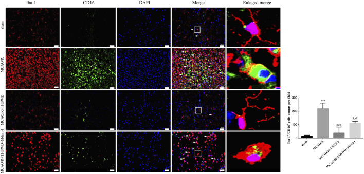
Globally, cerebral ischemia has been shown to be the second leading cause of death. Our previous studies have shown that Taohong Siwu Decoction (THSWD) exhibits obvious neuroprotective effects on cerebral ischemia/reperfusion (I/R) injury (CIRI). In this study, we further explored the modulatory effect of THSWD on mitochondrial autophagy in CIRI and the relationship between modulatory effect and NLRP3 inflammatory vesicle activation, so as to further explain the mechanism of neuroprotective effect of THSWD. Middle cerebral artery occlusion reperfusion (MCAO/R) model in rats was built to simulate I/R. Adult male SD rats (220-270 g) were randomly divided into the following four groups: the sham group, the MCAO/R group, the MCAO/R + THSWD group, and the MCAO/R + THSWD + Mitochondrial division inhibitor 1 (Mdivi-1) group. Neurological defect scores were used to evaluate neurological function. 2,3,5-Triphenyltetrazolium chloride (TTC) staining was conducted to measure cerebral infarct volume. Nissl staining, H&E staining and TUNEL staining were executed to detect ischemic cortical neuronal cell viability and apoptosis. Electron microscopy was used to observe the ultrastructural changes of mitochondria. Total Reactive Oxygen Species (ROS) in tissue were measured by fluorescence spectrophotometry, and the activation status of microglia was evaluated by Iba-1/CD16 immunofluorescence staining. The levels of mitophagy-related proteins (LC3, Parkin, PINK1), NLRP3 inflammasome-related proteins (NLRP3, ASC, Pro-caspase-1, Cleaved-caspase-1), and inflammatory cytokines (Pro-IL-18, Pro-IL-1β, IL-18, IL-1β) were evaluated by western blotting. The studies showed that THSWD treatment alleviated cerebral infarction and neurological deficiencies. THSWD upregulated the expressions of autophagy markers (LC3-II/LC3-I and Beclin1) mitochondrial autophagy markers (Parkin and PINK1) after CIRI. Furthermore, THSWD treatment attenuated microglia activation and damage to mitochondrial structures, thereby reducing ROS production and NLRP3 inflammasome activation. In contrast, the mitochondrial autophagy inhibitor Mdivi-1 inhibited the above beneficial effects of THSWD. THSWD exhibits neuroprotective effects against MCAO/R in rats by enhancing mitochondrial autophagy and reducing NLRP3 inflammasome activation.
在全球范围内,脑缺血已被证明是第二大致死原因。我们之前的研究表明,桃红四物汤(THSWD)对脑缺血/再灌注(I/R)损伤(CIRI)具有明显的神经保护作用。在本研究中,我们进一步探讨了THSWD对CIRI中线粒体自噬的调节作用以及调节作用与NLRP3炎性小体激活之间的关系,以进一步解释THSWD神经保护作用的机制。建立大鼠大脑中动脉闭塞再灌注(MCAO/R)模型以模拟I/R。将成年雄性SD大鼠(220 - 270 g)随机分为以下四组:假手术组、MCAO/R组、MCAO/R + THSWD组和MCAO/R + THSWD +线粒体分裂抑制剂1(Mdivi-1)组。采用神经功能缺损评分评估神经功能。进行2,3,5 - 氯化三苯基四氮唑(TTC)染色以测量脑梗死体积。进行尼氏染色、苏木精 - 伊红(H&E)染色和TUNEL染色以检测缺血皮质神经元细胞的活力和凋亡。采用电子显微镜观察线粒体的超微结构变化。通过荧光分光光度法测量组织中的总活性氧(ROS),并通过Iba-1/CD16免疫荧光染色评估小胶质细胞的激活状态。通过蛋白质免疫印迹法评估线粒体自噬相关蛋白(LC3、Parkin、PINK1)、NLRP3炎性小体相关蛋白(NLRP3、ASC、前半胱天冬酶 - 1、裂解的半胱天冬酶 - 1)和炎性细胞因子(前白细胞介素 - 18、前白细胞介素 - 1β、白细胞介素 - 18、白细胞介素 - 1β)的水平。研究表明,THSWD治疗可减轻脑梗死和神经功能缺陷。THSWD上调了CIRI后自噬标志物(LC3-II/LC3-I和Beclin1)和线粒体自噬标志物(Parkin和PINK1)的表达。此外,THSWD治疗减弱了小胶质细胞的激活和线粒体结构的损伤,从而减少了ROS的产生和NLRP3炎性小体的激活。相比之下,线粒体自噬抑制剂Mdivi-1抑制了THSWD的上述有益作用。THSWD通过增强线粒体自噬和减少NLRP3炎性小体激活,对大鼠MCAO/R具有神经保护作用。