• 文献检索
  • 文档翻译
  • 深度研究
  • 学术资讯
  • Suppr Zotero 插件Zotero 插件
  • 邀请有礼
  • 套餐&价格
  • 历史记录
应用&插件
Suppr Zotero 插件Zotero 插件浏览器插件Mac 客户端Windows 客户端微信小程序
定价
高级版会员购买积分包购买API积分包
服务
文献检索文档翻译深度研究API 文档MCP 服务
关于我们
关于 Suppr公司介绍联系我们用户协议隐私条款
关注我们

Suppr 超能文献

核心技术专利:CN118964589B侵权必究
粤ICP备2023148730 号-1Suppr @ 2026

文献检索

告别复杂PubMed语法,用中文像聊天一样搜索,搜遍4000万医学文献。AI智能推荐,让科研检索更轻松。

立即免费搜索

文件翻译

保留排版,准确专业,支持PDF/Word/PPT等文件格式,支持 12+语言互译。

免费翻译文档

深度研究

AI帮你快速写综述,25分钟生成高质量综述,智能提取关键信息,辅助科研写作。

立即免费体验

三维结构光照明显微镜,轴向分辨率增强。

Three-dimensional structured illumination microscopy with enhanced axial resolution.

机构信息

Laboratory of High Resolution Optical Imaging, National Institute of Biomedical Imaging and Bioengineering, National Institutes of Health, Bethesda, MD, USA.

Janelia Research Campus, Howard Hughes Medical Institute (HHMI), Ashburn, VA, USA.

出版信息

Nat Biotechnol. 2023 Sep;41(9):1307-1319. doi: 10.1038/s41587-022-01651-1. Epub 2023 Jan 26.

DOI:10.1038/s41587-022-01651-1
PMID:36702897
原文链接:https://pmc.ncbi.nlm.nih.gov/articles/PMC10497409/
Abstract

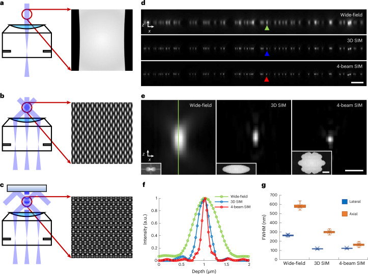
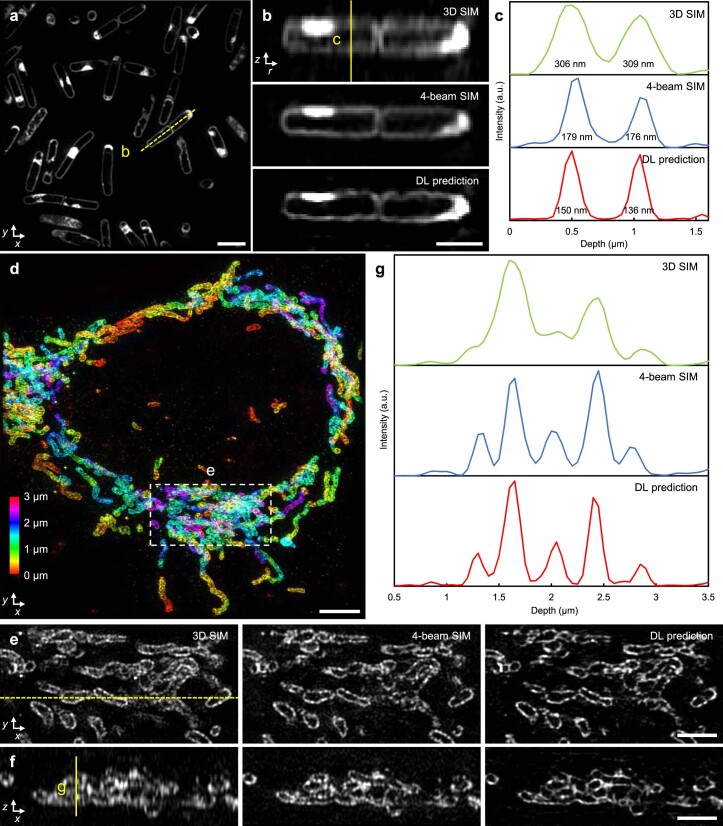
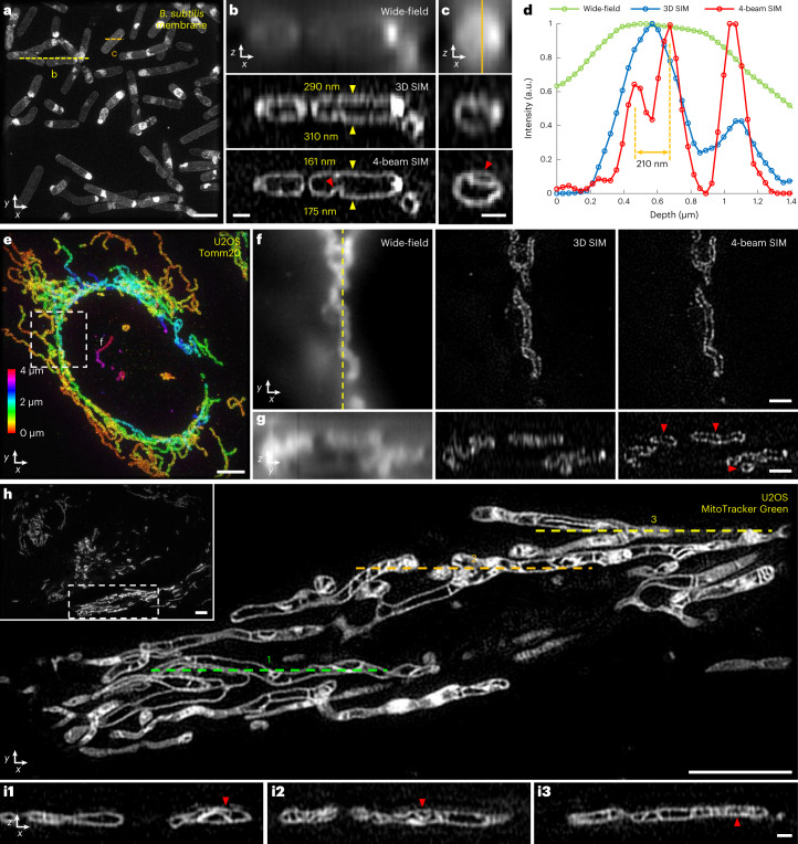
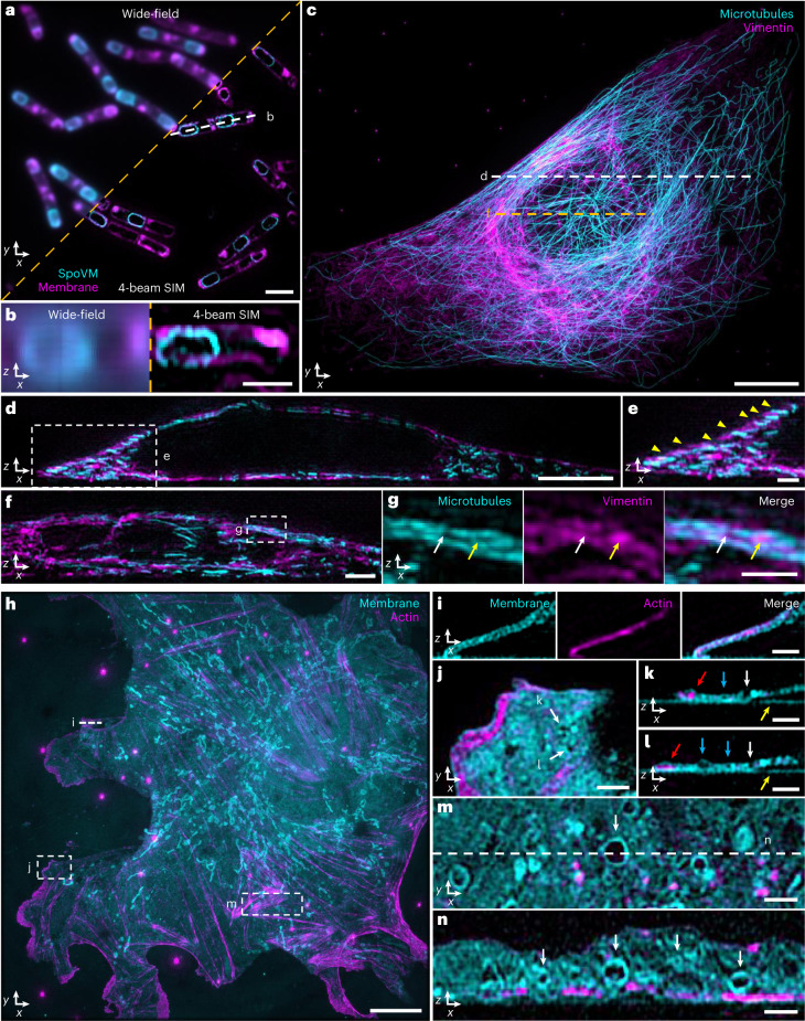
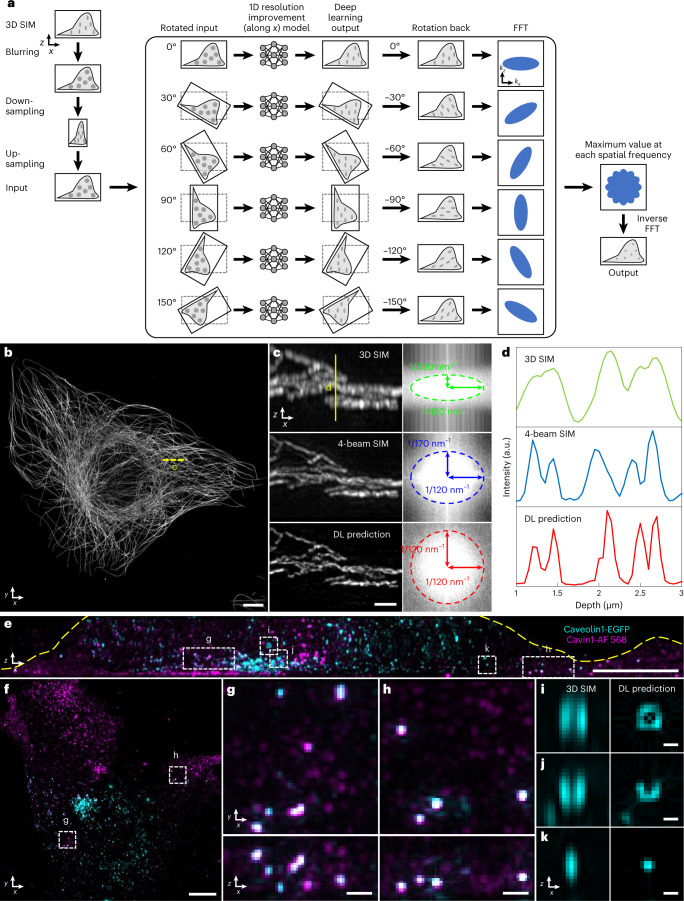
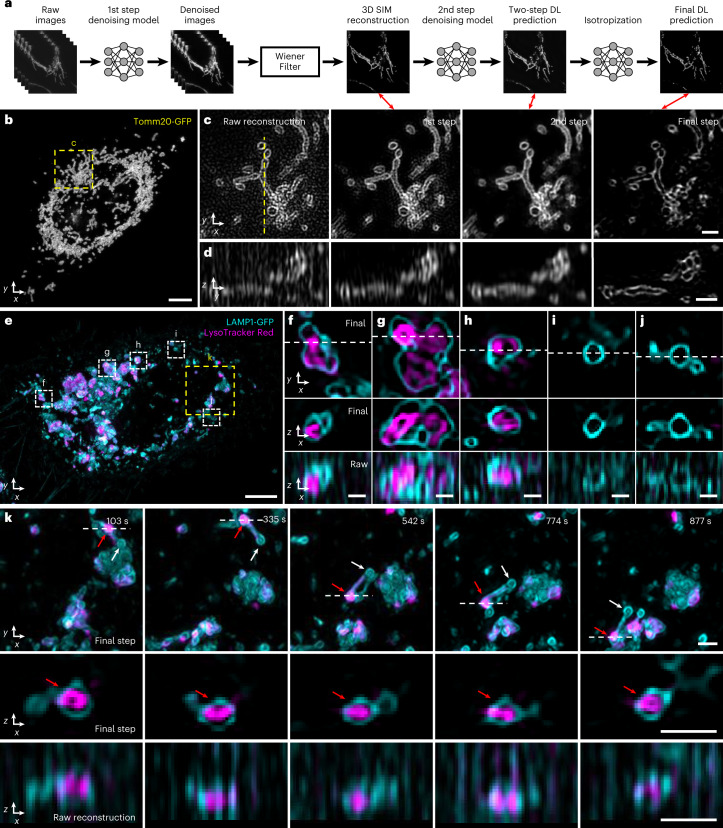
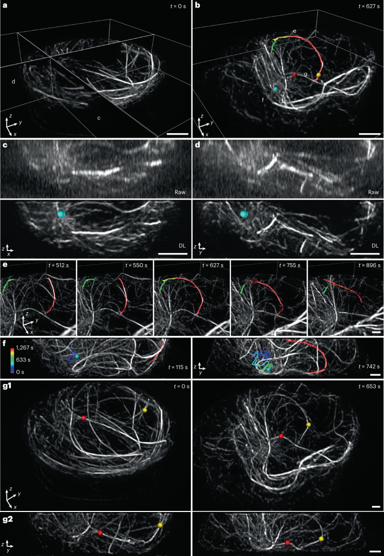
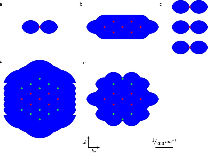
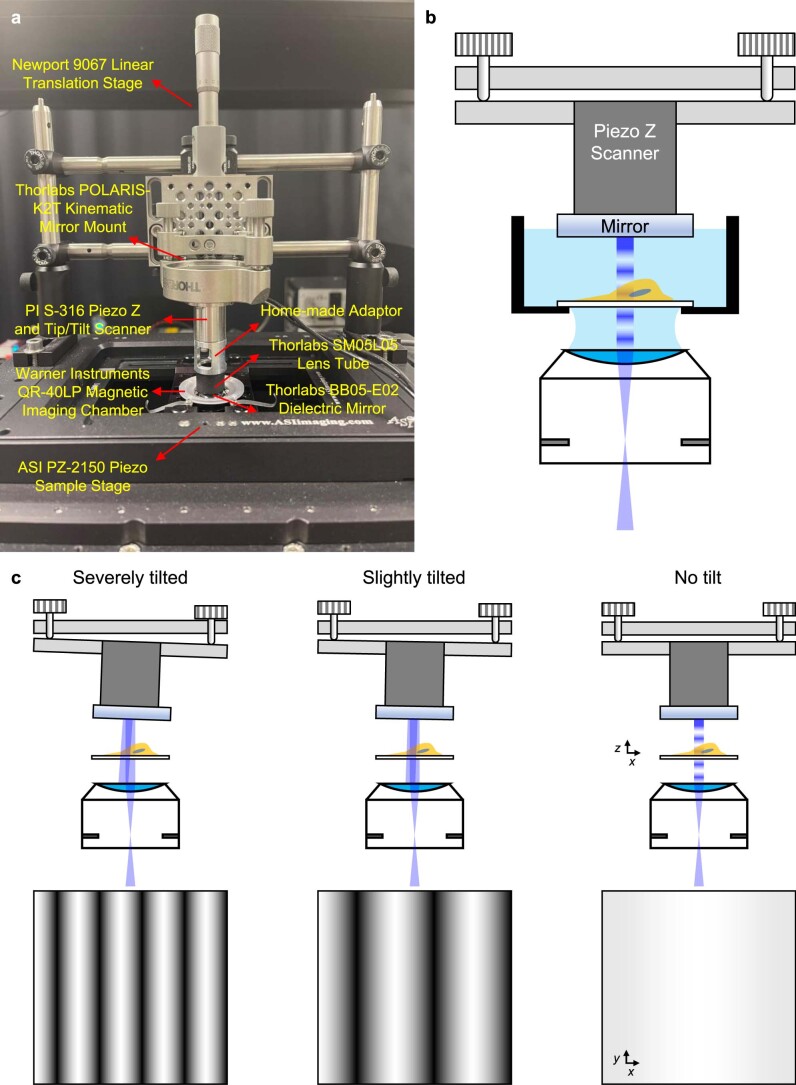
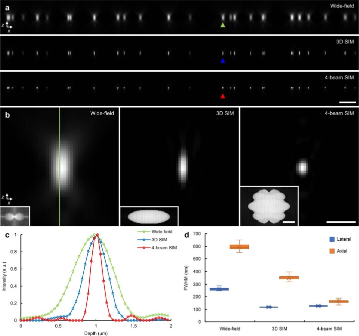
The axial resolution of three-dimensional structured illumination microscopy (3D SIM) is limited to ∼300 nm. Here we present two distinct, complementary methods to improve axial resolution in 3D SIM with minimal or no modification to the optical system. We show that placing a mirror directly opposite the sample enables four-beam interference with higher spatial frequency content than 3D SIM illumination, offering near-isotropic imaging with ∼120-nm lateral and 160-nm axial resolution. We also developed a deep learning method achieving ∼120-nm isotropic resolution. This method can be combined with denoising to facilitate volumetric imaging spanning dozens of timepoints. We demonstrate the potential of these advances by imaging a variety of cellular samples, delineating the nanoscale distribution of vimentin and microtubule filaments, observing the relative positions of caveolar coat proteins and lysosomal markers and visualizing cytoskeletal dynamics within T cells in the early stages of immune synapse formation.

摘要

三维结构光照明显微镜(3D SIM)的轴向分辨率限制在约 300nm。在这里,我们提出了两种不同的、互补的方法,通过对光学系统进行最小或不修改来提高 3D SIM 中的轴向分辨率。我们表明,在样品的正对面放置一面镜子,可以实现具有比 3D SIM 照明更高空间频率内容的四束干涉,从而提供具有约 120nm 侧向和 160nm 轴向分辨率的各向同性成像。我们还开发了一种深度学习方法,实现了约 120nm 的各向同性分辨率。该方法可以与去噪相结合,以促进数十个时间点的体积成像。我们通过对各种细胞样本进行成像来证明这些进展的潜力,描绘出中间丝和微管丝的纳米尺度分布,观察网格蛋白包被蛋白和溶酶体标记物的相对位置,并观察免疫突触形成早期 T 细胞内细胞骨架动力学。

https://cdn.ncbi.nlm.nih.gov/pmc/blobs/7047/10497409/93b2a25c3dbe/41587_2022_1651_Fig10_ESM.jpg
https://cdn.ncbi.nlm.nih.gov/pmc/blobs/7047/10497409/bb2237834371/41587_2022_1651_Fig1_HTML.jpg
https://cdn.ncbi.nlm.nih.gov/pmc/blobs/7047/10497409/97ac00f1dba2/41587_2022_1651_Fig2_HTML.jpg
https://cdn.ncbi.nlm.nih.gov/pmc/blobs/7047/10497409/647ab8f56115/41587_2022_1651_Fig3_HTML.jpg
https://cdn.ncbi.nlm.nih.gov/pmc/blobs/7047/10497409/b5b1fc5bf275/41587_2022_1651_Fig4_HTML.jpg
https://cdn.ncbi.nlm.nih.gov/pmc/blobs/7047/10497409/151abefa01ab/41587_2022_1651_Fig5_HTML.jpg
https://cdn.ncbi.nlm.nih.gov/pmc/blobs/7047/10497409/552108903745/41587_2022_1651_Fig6_HTML.jpg
https://cdn.ncbi.nlm.nih.gov/pmc/blobs/7047/10497409/a8e2b5993b68/41587_2022_1651_Fig7_ESM.jpg
https://cdn.ncbi.nlm.nih.gov/pmc/blobs/7047/10497409/e63833c0e264/41587_2022_1651_Fig8_ESM.jpg
https://cdn.ncbi.nlm.nih.gov/pmc/blobs/7047/10497409/9434552627d8/41587_2022_1651_Fig9_ESM.jpg
https://cdn.ncbi.nlm.nih.gov/pmc/blobs/7047/10497409/93b2a25c3dbe/41587_2022_1651_Fig10_ESM.jpg

相似文献

1
Three-dimensional structured illumination microscopy with enhanced axial resolution.三维结构光照明显微镜,轴向分辨率增强。
Nat Biotechnol. 2023 Sep;41(9):1307-1319. doi: 10.1038/s41587-022-01651-1. Epub 2023 Jan 26.
2
Resolution doubling in light-sheet microscopy via oblique plane structured illumination.利用斜平面结构照明实现光片显微镜的分辨率倍增。
Nat Methods. 2022 Nov;19(11):1419-1426. doi: 10.1038/s41592-022-01635-8. Epub 2022 Oct 24.
3
Three-dimensional super-resolution imaging of live whole cells using galvanometer-based structured illumination microscopy.使用基于振镜的结构照明显微镜对活的完整细胞进行三维超分辨率成像。
Opt Express. 2019 Mar 4;27(5):7237-7248. doi: 10.1364/OE.27.007237.
4
Super-Resolution Imaging of Plasmodesmata Using 3D Structured Illumination Microscopy.利用三维结构光照明显微镜对胞间连丝进行超分辨率成像。
Methods Mol Biol. 2022;2457:143-148. doi: 10.1007/978-1-0716-2132-5_8.
5
MAxSIM: multi-angle-crossing structured illumination microscopy with height-controlled mirror for 3D topological mapping of live cells.MAxSIM:具有高度可控反射镜的多角度交叉结构照明显微镜,用于活细胞的 3D 拓扑映射。
Commun Biol. 2023 Oct 12;6(1):1034. doi: 10.1038/s42003-023-05380-2.
6
Subcellular three-dimensional imaging deep through multicellular thick samples by structured illumination microscopy and adaptive optics.基于结构光照明显微镜和自适应光学的三维亚细胞深层成像技术在多细胞厚样本中的应用
Nat Commun. 2021 May 25;12(1):3148. doi: 10.1038/s41467-021-23449-6.
7
Recent advancements in structured-illumination microscopy toward live-cell imaging.结构照明显微镜在活细胞成像方面的最新进展。
Microscopy (Oxf). 2015 Aug;64(4):237-49. doi: 10.1093/jmicro/dfv034. Epub 2015 Jun 30.
8
Improving axial resolution in Structured Illumination Microscopy using deep learning.利用深度学习提高结构光照明显微镜的轴向分辨率。
Philos Trans A Math Phys Eng Sci. 2021 Jun 14;379(2199):20200298. doi: 10.1098/rsta.2020.0298. Epub 2021 Apr 26.
9
Correlative 3D Structured Illumination Microscopy and Single-Molecule Localization Microscopy for Imaging Cancer Invasion.用于成像癌症侵袭的相关三维结构光照明显微镜和单分子定位显微镜
Methods Mol Biol. 2018;1764:253-265. doi: 10.1007/978-1-4939-7759-8_15.
10
Super-resolution 3D microscopy of live whole cells using structured illumination.使用结构光照明显微镜对活的整个细胞进行超高分辨率 3D 成像。
Nat Methods. 2011 Oct 16;8(12):1044-6. doi: 10.1038/nmeth.1734.

引用本文的文献

1
Principal component analysis for three-dimensional structured illumination microscopy (PCA-3DSIM).用于三维结构光照显微镜的主成分分析(PCA-3DSIM)。
Light Sci Appl. 2025 Sep 1;14(1):299. doi: 10.1038/s41377-025-01979-8.
2
Adaptive-learning physics-assisted light-field microscopy enables day-long and millisecond-scale super-resolution imaging of 3D subcellular dynamics.自适应学习物理辅助光场显微镜实现了对三维亚细胞动力学的全天和毫秒级超分辨率成像。
Nat Commun. 2025 Aug 4;16(1):7132. doi: 10.1038/s41467-025-62471-w.
3
Extrachromosomal circular DNA drives dynamic genome plasticity: emerging roles in disease progression and clinical potential.

本文引用的文献

1
Incorporating the image formation process into deep learning improves network performance.将图像形成过程纳入深度学习可以提高网络性能。
Nat Methods. 2022 Nov;19(11):1427-1437. doi: 10.1038/s41592-022-01652-7. Epub 2022 Oct 31.
2
Multiview confocal super-resolution microscopy.多视场共聚焦超分辨显微镜。
Nature. 2021 Dec;600(7888):279-284. doi: 10.1038/s41586-021-04110-0. Epub 2021 Nov 26.
3
Volumetric interferometric lattice light-sheet imaging.体干涉格子光片成像。
染色体外环状DNA驱动动态基因组可塑性:在疾病进展中的新作用及临床潜力
Theranostics. 2025 May 25;15(13):6387-6411. doi: 10.7150/thno.111765. eCollection 2025.
4
Fast-adaptive super-resolution lattice light-sheet microscopy for rapid, long-term, near-isotropic subcellular imaging.用于快速、长期、近各向同性亚细胞成像的快速自适应超分辨率晶格光片显微镜。
Nat Methods. 2025 May;22(5):1059-1069. doi: 10.1038/s41592-025-02678-3. Epub 2025 Apr 29.
5
Computational Super-Resolution: An Odyssey in Harnessing Priors to Enhance Optical Microscopy Resolution.计算超分辨率:利用先验知识提升光学显微镜分辨率的探索之旅。
Anal Chem. 2025 Mar 11;97(9):4763-4792. doi: 10.1021/acs.analchem.4c07047. Epub 2025 Feb 27.
6
System- and sample-agnostic isotropic three-dimensional microscopy by weakly physics-informed, domain-shift-resistant axial deblurring.通过弱物理信息、抗域偏移轴向去模糊实现的与系统和样本无关的各向同性三维显微镜技术。
Nat Commun. 2025 Jan 16;16(1):745. doi: 10.1038/s41467-025-56078-4.
7
An in vivo CRISPR screen in chick embryos reveals a role for MLLT3 in specification of neural cells from the caudal epiblast.一项在鸡胚中的体内CRISPR筛选揭示了MLLT3在尾侧上胚层神经细胞特化中的作用。
Development. 2025 Feb 1;152(3). doi: 10.1242/dev.204591. Epub 2025 Feb 12.
8
Deep learning-based aberration compensation improves contrast and resolution in fluorescence microscopy.基于深度学习的像差补偿提高了荧光显微镜的对比度和分辨率。
Nat Commun. 2025 Jan 2;16(1):313. doi: 10.1038/s41467-024-55267-x.
9
Optical sectioning methods in three-dimensional bioimaging.三维生物成像中的光学切片方法。
Light Sci Appl. 2025 Jan 1;14(1):11. doi: 10.1038/s41377-024-01677-x.
10
Elucidating subcellular architecture and dynamics at isotropic 100-nm resolution with 4Pi-SIM.利用4Pi-SIM以各向同性100纳米分辨率阐明亚细胞结构和动力学。
Nat Methods. 2025 Feb;22(2):335-347. doi: 10.1038/s41592-024-02515-z. Epub 2024 Dec 23.
Nat Biotechnol. 2021 Nov;39(11):1385-1393. doi: 10.1038/s41587-021-01042-y. Epub 2021 Oct 11.
4
ML-SIM: universal reconstruction of structured illumination microscopy images using transfer learning.ML-SIM:基于迁移学习的结构光照明显微镜图像通用重建方法
Biomed Opt Express. 2021 Apr 15;12(5):2720-2733. doi: 10.1364/BOE.414680. eCollection 2021 May 1.
5
Time-resolved structured illumination microscopy reveals key principles of Xist RNA spreading.时间分辨结构照明显微术揭示了 Xist RNA 扩散的关键原则。
Science. 2021 Jun 11;372(6547). doi: 10.1126/science.abe7500.
6
Three-dimensional residual channel attention networks denoise and sharpen fluorescence microscopy image volumes.三维残差通道注意力网络对荧光显微镜图像体积进行降噪和锐化。
Nat Methods. 2021 Jun;18(6):678-687. doi: 10.1038/s41592-021-01155-x. Epub 2021 May 31.
7
Energy and Dynamics of Caveolae Trafficking.小窝运输的能量与动力学
Front Cell Dev Biol. 2021 Jan 21;8:614472. doi: 10.3389/fcell.2020.614472. eCollection 2020.
8
Dynein self-organizes while translocating the centrosome in T-cells.动力蛋白在 T 细胞中转运中心体时自我组织。
Mol Biol Cell. 2021 Apr 19;32(9):855-868. doi: 10.1091/mbc.E20-10-0668. Epub 2021 Mar 10.
9
Evaluation and development of deep neural networks for image super-resolution in optical microscopy.光学显微镜图像超分辨率的深度神经网络评估与发展。
Nat Methods. 2021 Feb;18(2):194-202. doi: 10.1038/s41592-020-01048-5. Epub 2021 Jan 21.
10
Volumetric live cell imaging with three-dimensional parallelized RESOLFT microscopy.三维平行 RESOLFT 显微镜的容积活细胞成像。
Nat Biotechnol. 2021 May;39(5):609-618. doi: 10.1038/s41587-020-00779-2. Epub 2021 Jan 11.