Ma Rui-Fang, Pao Ping-Chieh, Zhang Kun, Liu Jin-Xiang, Zhang Lin
Institute of Neuroscience Kunming Medical University Kunming Yunnan China.
Picower Institute for Learning and Memory, Department of Brain and Cognitive Sciences Massachusetts Institute of Technology Cambridge Massachusetts USA.
Ibrain. 2023 May 10;9(4):359-368. doi: 10.1002/ibra.12103. eCollection 2023 Winter.
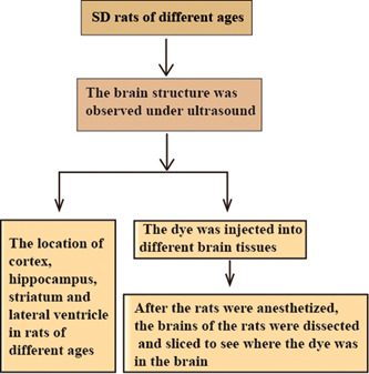
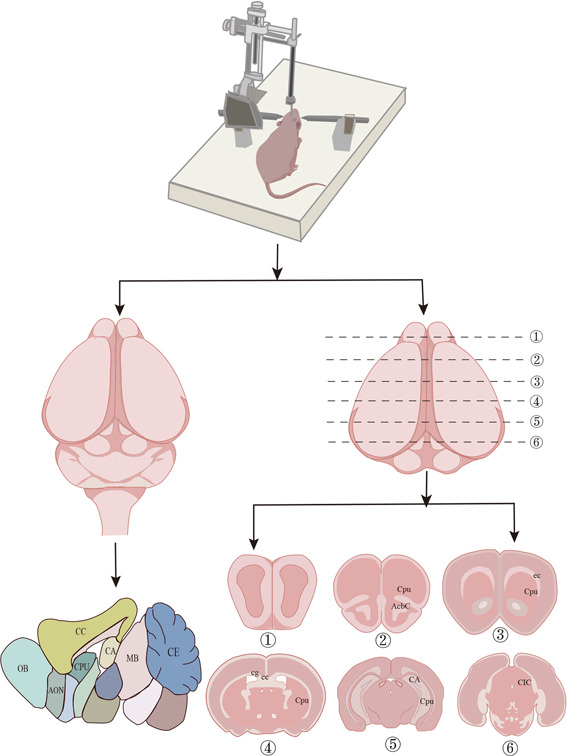
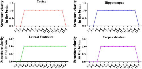
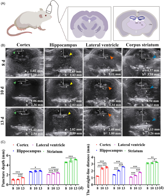
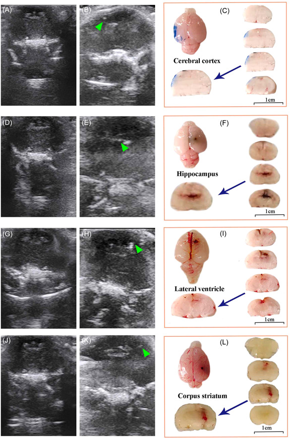
Since the brain structure of neonatal rats was not fully formed during the first 4 days, it cannot be detected using ultrasound. The objective of this study was to investigate the use of ultrasound to guide puncture in the normal coronal brain structure and determine the puncture depth of the location of the cortex, hippocampus, lateral ventricle, and striatum of newborn rats of 5-15 days. The animal was placed in a prone position. The specific positions of the cortex, hippocampus, lateral ventricle, and striatum were measured under ultrasound. Then, the rats were punctured with a stereotaxic instrument, and dye was injected. Finally, the brains of rats were taken to make frozen sections to observe the puncture results. By ultrasound, the image of the cortex, hippocampus, lateral ventricle, and striatum of the rat can be obtained and the puncture depth of the cortex (8 days: 1.02 ± 0.12, 10 days: 1.02 ± 0.08, 13 days: 1.43 ± 0.05), hippocampus (8 days: 2.63 ± 0.07, 10 days: 2.77 ± 0.14, 13 days: 2.82 ± 0.09), lateral ventricle (8 days: 2.08 ± 0.04, 10 days: 2.26 ± 0.03, 13 days: 2.40 ± 0.06), and corpus striatum (8 days: 4.57 ± 0.09, 10 days: 4.94 ± 0.31, 13 days: 5.13 ± 0.10) can be accurately measured. The rat brain structure and puncture depth changed with the age of the rats. Ultrasound technology can not only clarify the brain structure characteristics of 5-15-day-old rats but also guide the puncture and injection of the rat brain structure. The results of this study laid the foundation for the future use of ultrasound in experimental animal models of neurological diseases.
由于新生大鼠的脑结构在出生后的前4天尚未完全形成,因此无法用超声检测。本研究的目的是探讨超声引导下对正常冠状脑结构进行穿刺,并确定5 - 15日龄新生大鼠皮质、海马、侧脑室和纹状体部位的穿刺深度。将动物置于俯卧位。在超声下测量皮质、海马、侧脑室和纹状体的具体位置。然后,用立体定位仪对大鼠进行穿刺,并注入染料。最后,取出大鼠大脑制作冰冻切片以观察穿刺结果。通过超声,可以获得大鼠皮质、海马、侧脑室和纹状体的图像,并且可以准确测量皮质(8日龄:1.02±0.12,10日龄:1.02±0.08,13日龄:1.43±0.05)、海马(8日龄:2.63±0.07,10日龄:2.77±0.14,13日龄:2.82±0.09)、侧脑室(8日龄:2.08±0.04,10日龄:2.26±0.03,13日龄:2.40±0.06)和纹状体(8日龄:4.57±0.09,10日龄:4.94±0.31,13日龄:5.13±0.10)的穿刺深度。大鼠脑结构和穿刺深度随大鼠年龄而变化。超声技术不仅可以明确5 - 15日龄大鼠的脑结构特征,还可以指导对大鼠脑结构进行穿刺和注射。本研究结果为未来超声在神经疾病实验动物模型中的应用奠定了基础。