Campos Lillian Gonçalves, de Oliveira Francine Hehn, Antunes Ápio Cláudio Martins, Duarte Juliana Ávila
Department of Radiology, Hospital de Clínicas de Porto Alegre (HCPA), Porto Alegre, RS, Brazil.
Universidade Federal do Rio Grande do Sul (UFRGS), Porto Alegre, RS, Brazil.
Radiol Bras. 2024 Sep 16;57:e20240025. doi: 10.1590/0100-3984.2024.0025. eCollection 2024 Jan-Dec.
To determine the correlation of conventional and diffusion-weighted imaging findings on magnetic resonance imaging (MRI) of the brain, based on Visually AcceSAble Rembrandt Images (VASARI) criteria, with the histopathological grading of gliomas: low-grade or high-grade.
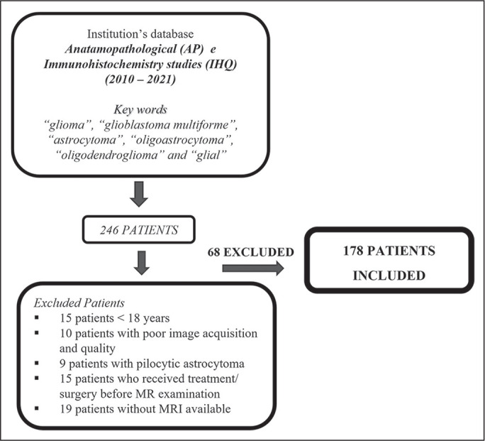
Preoperative MRI scans of 178 patients with brain gliomas and pathological confirmation were rated by two neuroradiologists for tumor size, location, and tumor morphology, using a standardized imaging feature set based on the VASARI criteria.
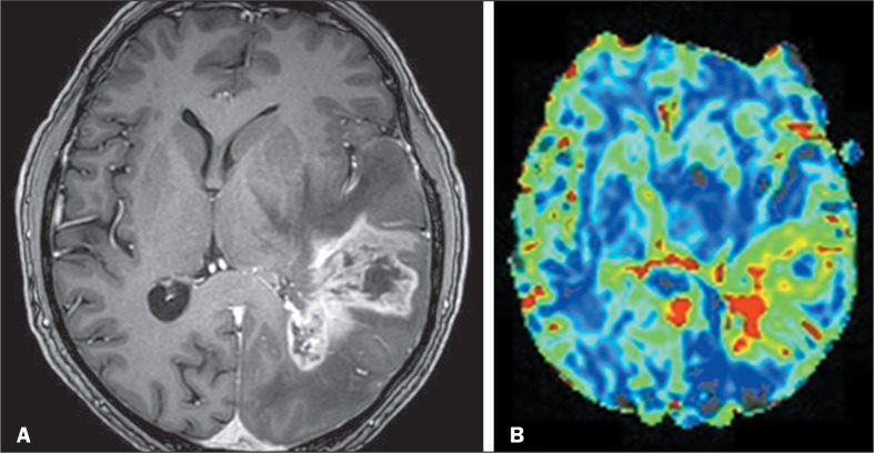
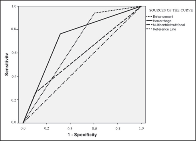
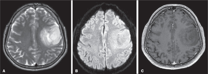
In the univariate analysis, more than half of the MRI characteristics evaluated showed a significant association with the tumor grade. The characteristics most significantly associated with the tumor grade were hemorrhage; restricted diffusion; pial invasion; enhancement; and a non-contrast-enhancing tumor crossing the midline. In a multivariable regression model, the presence of enhancement and hemorrhage maintained a significant association with high tumor grade. The absence of contrast enhancement and restricted diffusion were associated with the presence of an isocitrate dehydrogenase gene mutation.
Our data illustrate that VASARI MRI features, especially intratumoral hemorrhage, contrast enhancement, and multicentricity, correlate strongly with glial tumor grade.
基于视觉可及的伦勃朗图像(VASARI)标准,确定脑磁共振成像(MRI)上常规成像和扩散加权成像结果与胶质瘤组织病理学分级(低级别或高级别)之间的相关性。
178例脑胶质瘤患者的术前MRI扫描及病理确诊结果由两名神经放射科医生根据基于VASARI标准的标准化成像特征集对肿瘤大小、位置和肿瘤形态进行评估。
在单变量分析中,所评估的MRI特征中超过一半与肿瘤分级存在显著关联。与肿瘤分级最显著相关的特征为出血;扩散受限;软脑膜侵犯;强化;以及跨越中线的无强化肿瘤。在多变量回归模型中,强化和出血的存在与高级别肿瘤仍保持显著关联。无强化和扩散受限与异柠檬酸脱氢酶基因突变的存在相关。
我们的数据表明,VASARI MRI特征,尤其是肿瘤内出血、强化和多中心性,与胶质肿瘤分级密切相关。