• 文献检索
  • 文档翻译
  • 深度研究
  • 学术资讯
  • Suppr Zotero 插件Zotero 插件
  • 邀请有礼
  • 套餐&价格
  • 历史记录
应用&插件
Suppr Zotero 插件Zotero 插件浏览器插件Mac 客户端Windows 客户端微信小程序
定价
高级版会员购买积分包购买API积分包
服务
文献检索文档翻译深度研究API 文档MCP 服务
关于我们
关于 Suppr公司介绍联系我们用户协议隐私条款
关注我们

Suppr 超能文献

核心技术专利:CN118964589B侵权必究
粤ICP备2023148730 号-1Suppr @ 2026

文献检索

告别复杂PubMed语法,用中文像聊天一样搜索,搜遍4000万医学文献。AI智能推荐,让科研检索更轻松。

立即免费搜索

文件翻译

保留排版,准确专业,支持PDF/Word/PPT等文件格式,支持 12+语言互译。

免费翻译文档

深度研究

AI帮你快速写综述,25分钟生成高质量综述,智能提取关键信息,辅助科研写作。

立即免费体验

利用肌动蛋白结合结构域与埃兹蛋白绿色荧光蛋白融合体对活的NIH3T3细胞微延伸中肌动蛋白细胞骨架的动态变化进行成像。

Imaging of dynamic changes of the actin cytoskeleton in microextensions of live NIH3T3 cells with a GFP fusion of the F-actin binding domain of moesin.

作者信息

Litman P, Amieva M R, Furthmayr H

机构信息

Molecular Mechanisms of Disease Laboratories, Department of Pathology, Stanford University Medical School, 300 Pasteur Drive, Stanford, CA 94305-5324, USA.

出版信息

BMC Cell Biol. 2000;1:1. doi: 10.1186/1471-2121-1-1. Epub 2000 Nov 1.

DOI:10.1186/1471-2121-1-1
PMID:11112983
原文链接:https://pmc.ncbi.nlm.nih.gov/articles/PMC29062/
Abstract

BACKGROUND

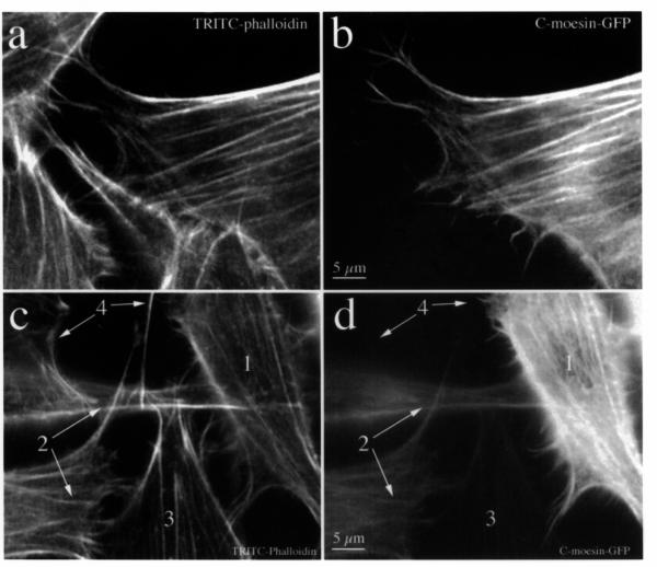
The cell surface undergoes continuous change during cell movement. This is characterized by transient protrusion and partial or complete retraction of microspikes, filopodia, and lamellipodia. This requires a dynamic actin cytoskeleton, moesin, components of Rho-mediated signal pathways, rearrangement of membrane constituents and the formation of focal adhesion sites. While the immunofluorescence distribution of endogenous moesin is that of a membrane-bound molecule with marked enhancement in some but not all microextensions, the C-terminal fragment of moesin co-distributes with filamentous actin consistent with its actin-binding activity. By taking advantage of this property we studied the spontaneous protrusive activity of live NIH3T3 cells, expressing a fusion of GFP and the C-terminal domain of moesin.

RESULTS

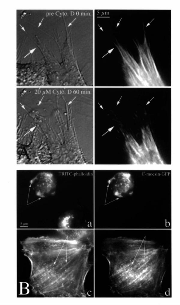
C-moesin-GFP localized to stress fibers and was enriched in actively protruding cellular regions such as filopodia or lamellipodia. This localization was reversibly affected by cytochalasin D. Multiple types of cytoskeletal rearrangements were observed that occurred independent of each other in adjacent regions of the cell surface. Assembly and disassembly of actin filaments occurred repeatedly within the same space and was correlated with either membrane protrusion and retraction, or no change in shape when microextensions were adherent.

CONCLUSIONS

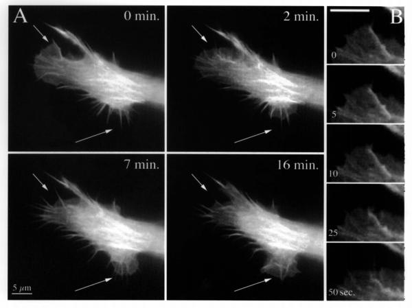
Shape alone provided an inadequate criterion for distinguishing between retraction fibers and advancing, retracting or stable filopodia. Fluorescence imaging of C-moesin-GFP, however, paralleled the rapid and dynamic changes of the actin cytoskeleton in microextensions. Regional regulatory control is implicated because opposite changes occurred in close proximity and presumably independent of each other. This new and sensitive tool should be useful for investigating mechanisms of localized actin dynamics in the cell cortex.

摘要

背景

细胞表面在细胞移动过程中会持续发生变化。其特征表现为微刺、丝状伪足和片状伪足的短暂突出以及部分或完全缩回。这需要动态的肌动蛋白细胞骨架、埃兹蛋白、Rho介导的信号通路成分、膜成分的重排以及粘着斑位点的形成。虽然内源性埃兹蛋白的免疫荧光分布是一种膜结合分子,在一些而非所有微延伸部位有明显增强,但埃兹蛋白的C末端片段与丝状肌动蛋白共分布,这与其肌动蛋白结合活性一致。利用这一特性,我们研究了表达绿色荧光蛋白(GFP)与埃兹蛋白C末端结构域融合蛋白的活NIH3T3细胞的自发突出活性。

结果

C-埃兹蛋白-GFP定位于应力纤维,并在诸如丝状伪足或片状伪足等活跃突出的细胞区域富集。这种定位受到细胞松弛素D的可逆影响。观察到多种类型的细胞骨架重排在细胞表面的相邻区域相互独立地发生。肌动蛋白丝的组装和拆卸在同一空间内反复发生,并且与膜的突出和缩回相关,或者当微延伸部位粘着时形状无变化。

结论

仅形状不足以作为区分缩回纤维与前进、缩回或稳定丝状伪足的标准。然而,C-埃兹蛋白-GFP的荧光成像与微延伸部位肌动蛋白细胞骨架的快速动态变化平行。由于在紧邻区域发生了相反的变化且可能相互独立,这暗示了区域调节控制。这种新的灵敏工具对于研究细胞皮层中局部肌动蛋白动力学机制应是有用的。

https://cdn.ncbi.nlm.nih.gov/pmc/blobs/4b89/29062/bf880955eda5/1471-2121-1-1-6.jpg
https://cdn.ncbi.nlm.nih.gov/pmc/blobs/4b89/29062/5618c8938b39/1471-2121-1-1-1.jpg
https://cdn.ncbi.nlm.nih.gov/pmc/blobs/4b89/29062/b71b7425d83b/1471-2121-1-1-2.jpg
https://cdn.ncbi.nlm.nih.gov/pmc/blobs/4b89/29062/00db9225a3b3/1471-2121-1-1-3.jpg
https://cdn.ncbi.nlm.nih.gov/pmc/blobs/4b89/29062/8ab724c787be/1471-2121-1-1-4.jpg
https://cdn.ncbi.nlm.nih.gov/pmc/blobs/4b89/29062/a240c2fa5e26/1471-2121-1-1-5.jpg
https://cdn.ncbi.nlm.nih.gov/pmc/blobs/4b89/29062/bf880955eda5/1471-2121-1-1-6.jpg

相似文献

1
Imaging of dynamic changes of the actin cytoskeleton in microextensions of live NIH3T3 cells with a GFP fusion of the F-actin binding domain of moesin.利用肌动蛋白结合结构域与埃兹蛋白绿色荧光蛋白融合体对活的NIH3T3细胞微延伸中肌动蛋白细胞骨架的动态变化进行成像。
BMC Cell Biol. 2000;1:1. doi: 10.1186/1471-2121-1-1. Epub 2000 Nov 1.
2
Disruption of dynamic cell surface architecture of NIH3T3 fibroblasts by the N-terminal domains of moesin and ezrin: in vivo imaging with GFP fusion proteins.埃兹蛋白(ezrin)和膜突蛋白(moesin)的N端结构域对NIH3T3成纤维细胞动态细胞表面结构的破坏:绿色荧光蛋白(GFP)融合蛋白的体内成像
J Cell Sci. 1999 Jan;112 ( Pt 1):111-25. doi: 10.1242/jcs.112.1.111.
3
Subcellular localization of moesin in dynamic filopodia, retraction fibers, and other structures involved in substrate exploration, attachment, and cell-cell contacts.膜突蛋白在动态丝状伪足、收缩纤维以及参与底物探索、附着和细胞间接触的其他结构中的亚细胞定位。
Exp Cell Res. 1995 Jul;219(1):180-96. doi: 10.1006/excr.1995.1218.
4
GFP-moesin illuminates actin cytoskeleton dynamics in living tissue and demonstrates cell shape changes during morphogenesis in Drosophila.绿色荧光蛋白标记的膜突蛋白可照亮活组织中的肌动蛋白细胞骨架动态变化,并揭示果蝇形态发生过程中的细胞形状变化。
Dev Biol. 1997 Nov 1;191(1):103-17. doi: 10.1006/dbio.1997.8707.
5
In vivo dynamics of the F-actin-binding protein neurabin-II.F-肌动蛋白结合蛋白神经素-II的体内动力学
Biochem J. 2000 Jan 15;345 Pt 2(Pt 2):185-94.
6
The suitability and application of a GFP-actin fusion protein for long-term imaging of the organization and dynamics of the cytoskeleton in mammalian cells.绿色荧光蛋白-肌动蛋白融合蛋白在哺乳动物细胞中对细胞骨架组织和动力学进行长期成像的适用性及应用
Eur J Cell Biol. 1998 Oct;77(2):81-90. doi: 10.1016/S0171-9335(98)80075-7.
7
Comparative analysis of tools for live cell imaging of actin network architecture.肌动蛋白网络结构活细胞成像工具的比较分析
Bioarchitecture. 2014;4(6):189-202. doi: 10.1080/19490992.2014.1047714. Epub 2015 Aug 28.
8
Differential localisation of GFP fusions to cytoskeleton-binding proteins in animal, plant, and yeast cells. Green-fluorescent protein.绿色荧光蛋白(GFP)融合蛋白在动物、植物和酵母细胞中与细胞骨架结合蛋白的差异定位。绿色荧光蛋白。
Protoplasma. 2002 Oct;220(1-2):69-78. doi: 10.1007/s00709-002-0026-7.
9
Moesin orchestrates cortical polarity of melanoma tumour cells to initiate 3D invasion.埃兹蛋白调控黑色素瘤肿瘤细胞的皮质极性以启动三维侵袭。
J Cell Sci. 2009 Oct 1;122(Pt 19):3492-501. doi: 10.1242/jcs.053157. Epub 2009 Sep 1.
10
Merlin differs from moesin in binding to F-actin and in its intra- and intermolecular interactions.Merlin在与F-肌动蛋白结合以及其分子内和分子间相互作用方面与埃兹蛋白不同。
Biochem Biophys Res Commun. 1998 Jul 30;248(3):548-53. doi: 10.1006/bbrc.1998.9009.

引用本文的文献

1
Tether-guided lamellipodia enable rapid wound healing.牵拉引导的片状伪足有助于快速伤口愈合。
Biophys J. 2022 Mar 15;121(6):1029-1037. doi: 10.1016/j.bpj.2022.02.006. Epub 2022 Feb 12.
2
Enhanced osseointegration through direct energy deposition porous coating for cementless orthopedic implant fixation.通过直接能量沉积多孔涂层增强骨整合,实现无水泥骨科植入物固定。
Sci Rep. 2021 Nov 16;11(1):22317. doi: 10.1038/s41598-021-01739-9.
3
Biomechanics and the thermotolerance of development.生物力学与发育的耐热性

本文引用的文献

1
Phosphatidylinositol 4,5-bisphosphate functions as a second messenger that regulates cytoskeleton-plasma membrane adhesion.磷脂酰肌醇4,5-二磷酸作为一种第二信使,调节细胞骨架与质膜的黏附。
Cell. 2000 Jan 21;100(2):221-8. doi: 10.1016/s0092-8674(00)81560-3.
2
Protein phosphatase 2C inactivates F-actin binding of human platelet moesin.蛋白磷酸酶2C使人类血小板埃兹蛋白的F-肌动蛋白结合失活。
J Biol Chem. 1999 Sep 17;274(38):26705-12. doi: 10.1074/jbc.274.38.26705.
3
Calcium influx alters actin bundle dynamics and retrograde flow in Helisoma growth cones.
PLoS One. 2014 Apr 28;9(4):e95670. doi: 10.1371/journal.pone.0095670. eCollection 2014.
4
Punctuated actin contractions during convergent extension and their permissive regulation by the non-canonical Wnt-signaling pathway.在趋同延伸过程中,肌动蛋白的间歇性收缩及其非经典 Wnt 信号通路的许可调节。
J Cell Sci. 2011 Feb 15;124(Pt 4):635-46. doi: 10.1242/jcs.067579. Epub 2011 Jan 25.
5
Macroscopic stiffening of embryonic tissues via microtubules, RhoGEF and the assembly of contractile bundles of actomyosin.通过微管、RhoGEF 和肌动球蛋白收缩束的组装使胚胎组织产生宏观僵硬。
Development. 2010 Aug;137(16):2785-94. doi: 10.1242/dev.045997. Epub 2010 Jul 14.
6
Dephosphorylation-dependent inhibitory activity of juxtanodin on filamentous actin disassembly.衔接蛋白聚糖对丝状肌动蛋白解聚的去磷酸化依赖性抑制活性。
J Biol Chem. 2010 Sep 10;285(37):28838-49. doi: 10.1074/jbc.M110.117887. Epub 2010 Jul 7.
7
Actin interaction and regulation of cyclin-dependent kinase 5/p35 complex activity.肌动蛋白的相互作用和细胞周期蛋白依赖性激酶 5/p35 复合物活性的调节。
J Neurochem. 2011 Jan;116(2):192-204. doi: 10.1111/j.1471-4159.2010.06824.x.
8
Experimental control of excitable embryonic tissues: three stimuli induce rapid epithelial contraction.实验控制可兴奋胚胎组织:三种刺激可快速诱导上皮细胞收缩。
Exp Cell Res. 2010 Jan 1;316(1):103-14. doi: 10.1016/j.yexcr.2009.08.005. Epub 2009 Aug 15.
9
Myosin II motors and F-actin dynamics drive the coordinated movement of the centrosome and soma during CNS glial-guided neuronal migration.肌球蛋白II马达和F-肌动蛋白动力学在中枢神经系统胶质细胞引导的神经元迁移过程中驱动中心体和细胞体的协调运动。
Neuron. 2009 Jul 16;63(1):63-80. doi: 10.1016/j.neuron.2009.05.028.
10
Morphogenetic movements driving neural tube closure in Xenopus require myosin IIB.非洲爪蟾中驱动神经管闭合的形态发生运动需要肌球蛋白IIB。
Dev Biol. 2009 Mar 15;327(2):327-38. doi: 10.1016/j.ydbio.2008.12.009. Epub 2008 Dec 24.
钙离子内流改变了扁卷螺生长锥中肌动蛋白束的动力学和逆向流动。
J Neurosci. 1999 Sep 15;19(18):7971-82. doi: 10.1523/JNEUROSCI.19-18-07971.1999.
4
Regulation of F-actin binding to platelet moesin in vitro by both phosphorylation of threonine 558 and polyphosphatidylinositides.苏氨酸558磷酸化和多磷脂酰肌醇对体外F-肌动蛋白与血小板埃兹蛋白结合的调节作用。
Mol Biol Cell. 1999 Aug;10(8):2669-85. doi: 10.1091/mbc.10.8.2669.
5
Induction of filopodia by direct local elevation of intracellular calcium ion concentration.通过直接局部提高细胞内钙离子浓度诱导丝状伪足形成。
J Cell Biol. 1999 Jun 14;145(6):1265-75. doi: 10.1083/jcb.145.6.1265.
6
Replacement of threonine 558, a critical site of phosphorylation of moesin in vivo, with aspartate activates F-actin binding of moesin. Regulation by conformational change.将苏氨酸558(体内埃兹蛋白磷酸化的关键位点)替换为天冬氨酸可激活埃兹蛋白与F-肌动蛋白的结合。通过构象变化进行调控。
J Biol Chem. 1999 Apr 30;274(18):12803-10. doi: 10.1074/jbc.274.18.12803.
7
Regulation of cortical structure by the ezrin-radixin-moesin protein family.埃兹蛋白-根蛋白-膜突蛋白家族对皮质结构的调控
Curr Opin Cell Biol. 1999 Feb;11(1):109-16. doi: 10.1016/s0955-0674(99)80013-1.
8
Phosphorylation of moesin by rho-associated kinase (Rho-kinase) plays a crucial role in the formation of microvilli-like structures.由Rho相关激酶(Rho激酶)介导的埃兹蛋白磷酸化在微绒毛样结构的形成中起关键作用。
J Biol Chem. 1998 Dec 25;273(52):34663-6. doi: 10.1074/jbc.273.52.34663.
9
Disruption of dynamic cell surface architecture of NIH3T3 fibroblasts by the N-terminal domains of moesin and ezrin: in vivo imaging with GFP fusion proteins.埃兹蛋白(ezrin)和膜突蛋白(moesin)的N端结构域对NIH3T3成纤维细胞动态细胞表面结构的破坏:绿色荧光蛋白(GFP)融合蛋白的体内成像
J Cell Sci. 1999 Jan;112 ( Pt 1):111-25. doi: 10.1242/jcs.112.1.111.
10
The suitability and application of a GFP-actin fusion protein for long-term imaging of the organization and dynamics of the cytoskeleton in mammalian cells.绿色荧光蛋白-肌动蛋白融合蛋白在哺乳动物细胞中对细胞骨架组织和动力学进行长期成像的适用性及应用
Eur J Cell Biol. 1998 Oct;77(2):81-90. doi: 10.1016/S0171-9335(98)80075-7.