• 文献检索
  • 文档翻译
  • 深度研究
  • 学术资讯
  • Suppr Zotero 插件Zotero 插件
  • 邀请有礼
  • 套餐&价格
  • 历史记录
应用&插件
Suppr Zotero 插件Zotero 插件浏览器插件Mac 客户端Windows 客户端微信小程序
定价
高级版会员购买积分包购买API积分包
服务
文献检索文档翻译深度研究API 文档MCP 服务
关于我们
关于 Suppr公司介绍联系我们用户协议隐私条款
关注我们

Suppr 超能文献

核心技术专利:CN118964589B侵权必究
粤ICP备2023148730 号-1Suppr @ 2026

文献检索

告别复杂PubMed语法,用中文像聊天一样搜索,搜遍4000万医学文献。AI智能推荐,让科研检索更轻松。

立即免费搜索

文件翻译

保留排版,准确专业,支持PDF/Word/PPT等文件格式,支持 12+语言互译。

免费翻译文档

深度研究

AI帮你快速写综述,25分钟生成高质量综述,智能提取关键信息,辅助科研写作。

立即免费体验

对活的上皮细胞中角蛋白中间丝动态特性的见解。

Insights into the dynamic properties of keratin intermediate filaments in living epithelial cells.

作者信息

Yoon K H, Yoon M, Moir R D, Khuon S, Flitney F W, Goldman R D

机构信息

Department of Cell and Molecular Biology, Northwestern University Medical School, Chicago, Illinois 60611.

出版信息

J Cell Biol. 2001 Apr 30;153(3):503-16. doi: 10.1083/jcb.153.3.503.

DOI:10.1083/jcb.153.3.503
PMID:11331302
原文链接:https://pmc.ncbi.nlm.nih.gov/articles/PMC2190576/
Abstract

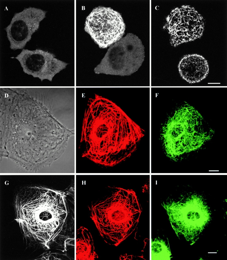
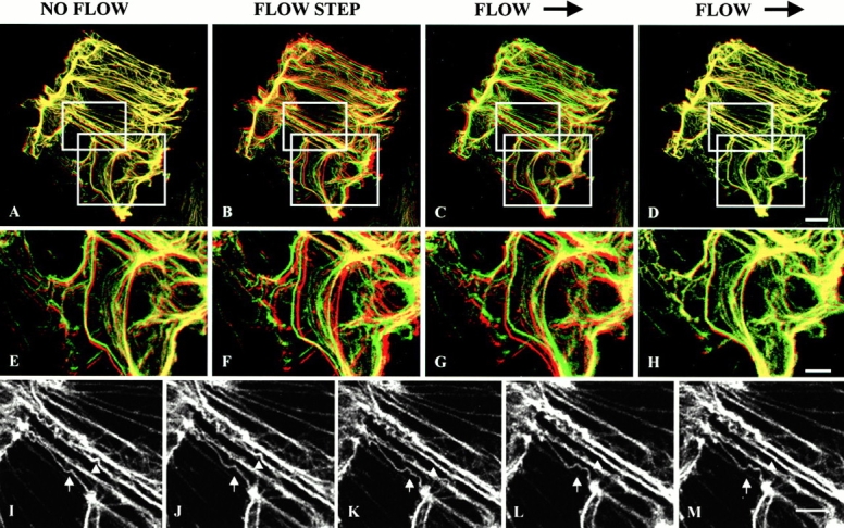
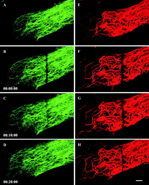
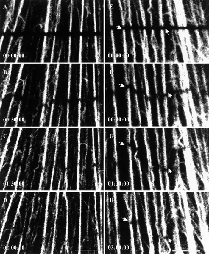
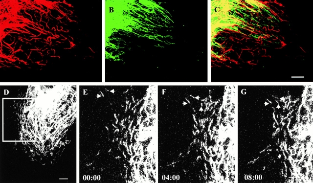
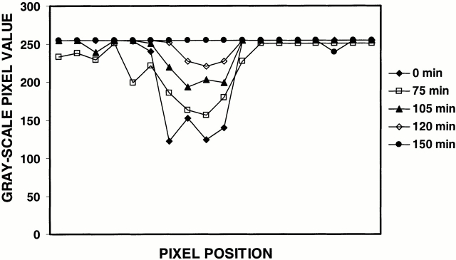
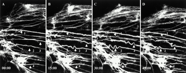
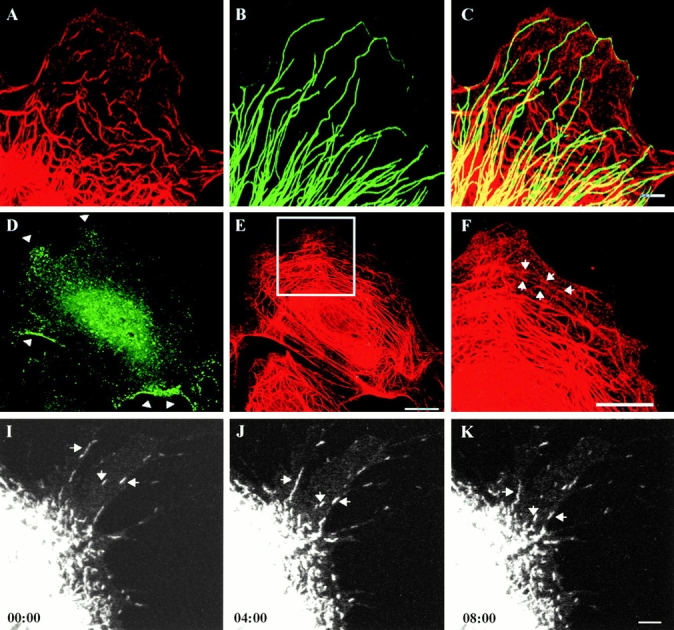
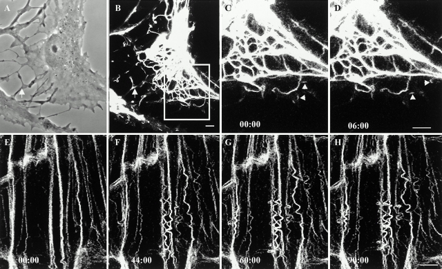
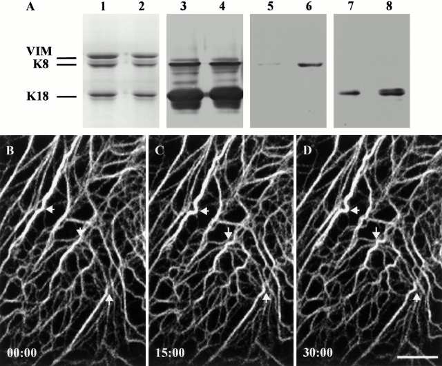
The properties of keratin intermediate filaments (IFs) have been studied after transfection with green fluorescent protein (GFP)-tagged K18 and/or K8 (type I/II IF proteins). GFP-K8 and -K18 become incorporated into tonofibrils, which are comprised of bundles of keratin IFs. These tonofibrils exhibit a remarkably wide range of motile and dynamic activities. Fluorescence recovery after photobleaching (FRAP) analyses show that they recover their fluorescence slowly with a recovery t(1/2) of approximately 100 min. The movements of bleach zones during recovery show that closely spaced tonofibrils (<1 microm apart) often move at different rates and in different directions. Individual tonofibrils frequently change their shapes, and in some cases these changes appear as propagated waveforms along their long axes. In addition, short fibrils, termed keratin squiggles, are seen at the cell periphery where they move mainly towards the cell center. The motile properties of keratin IFs are also compared with those of type III IFs (vimentin) in PtK2 cells. Intriguingly, the dynamic properties of keratin tonofibrils and squiggles are dramatically different from those of vimentin fibrils and squiggles within the same cytoplasmic regions. This suggests that there are different factors regulating the dynamic properties of different types of IFs within the same cytoplasmic regions.

摘要

在用绿色荧光蛋白(GFP)标记的K18和/或K8(I/II型中间丝蛋白)转染后,对角蛋白中间丝(IFs)的特性进行了研究。GFP-K8和-K18被整合到张力原纤维中,张力原纤维由角蛋白IFs束组成。这些张力原纤维表现出范围广泛的运动和动态活动。光漂白后荧光恢复(FRAP)分析表明,它们以约100分钟的恢复半衰期(t(1/2))缓慢恢复荧光。恢复过程中漂白区域的移动表明,紧密间隔的张力原纤维(间距<1微米)通常以不同速率和不同方向移动。单个张力原纤维经常改变其形状,在某些情况下,这些变化表现为沿其长轴传播的波形。此外,在细胞周边可见短纤维,称为角蛋白卷曲,它们主要向细胞中心移动。还将角蛋白IFs的运动特性与PtK2细胞中III型IFs(波形蛋白)的运动特性进行了比较。有趣的是,在同一细胞质区域内,角蛋白张力原纤维和卷曲的动态特性与波形蛋白纤维和卷曲的动态特性有显著差异。这表明在同一细胞质区域内,存在不同的因素调节不同类型IFs的动态特性。

https://cdn.ncbi.nlm.nih.gov/pmc/blobs/c9a6/2190576/785cbd4637d9/JCB0102001.f10.jpg
https://cdn.ncbi.nlm.nih.gov/pmc/blobs/c9a6/2190576/4339c710dcbd/JCB0102001.f1.jpg
https://cdn.ncbi.nlm.nih.gov/pmc/blobs/c9a6/2190576/3a473f0086f0/JCB0102001.f2.jpg
https://cdn.ncbi.nlm.nih.gov/pmc/blobs/c9a6/2190576/2439f2f60925/JCB0102001.f3.jpg
https://cdn.ncbi.nlm.nih.gov/pmc/blobs/c9a6/2190576/04e05a385e99/JCB0102001.f6.jpg
https://cdn.ncbi.nlm.nih.gov/pmc/blobs/c9a6/2190576/ccb00f2a7ad7/JCB0102001.f4.jpg
https://cdn.ncbi.nlm.nih.gov/pmc/blobs/c9a6/2190576/2e02bbe5f151/JCB0102001.f5.jpg
https://cdn.ncbi.nlm.nih.gov/pmc/blobs/c9a6/2190576/3df37cfbfa51/JCB0102001.f8.jpg
https://cdn.ncbi.nlm.nih.gov/pmc/blobs/c9a6/2190576/63a6f04df0a1/JCB0102001.f7.jpg
https://cdn.ncbi.nlm.nih.gov/pmc/blobs/c9a6/2190576/2d933dcf03ca/JCB0102001.f9.jpg
https://cdn.ncbi.nlm.nih.gov/pmc/blobs/c9a6/2190576/785cbd4637d9/JCB0102001.f10.jpg

相似文献

1
Insights into the dynamic properties of keratin intermediate filaments in living epithelial cells.对活的上皮细胞中角蛋白中间丝动态特性的见解。
J Cell Biol. 2001 Apr 30;153(3):503-16. doi: 10.1083/jcb.153.3.503.
2
Observation of keratin particles showing fast bidirectional movement colocalized with microtubules.观察到角蛋白颗粒呈现与微管共定位的快速双向运动。
J Cell Sci. 2003 Apr 15;116(Pt 8):1417-27. doi: 10.1242/jcs.00363.
3
Motile properties of vimentin intermediate filament networks in living cells.活细胞中波形蛋白中间丝网络的运动特性。
J Cell Biol. 1998 Oct 5;143(1):147-57. doi: 10.1083/jcb.143.1.147.
4
Insights into the mechanical properties of epithelial cells: the effects of shear stress on the assembly and remodeling of keratin intermediate filaments.上皮细胞力学特性的见解:剪切应力对角蛋白中间丝组装和重塑的影响。
FASEB J. 2009 Jul;23(7):2110-9. doi: 10.1096/fj.08-124453. Epub 2009 Feb 26.
5
Selective accumulation of IF proteins at a focal juxtanuclear site in COS-1 cells transfected with mouse keratin 18 cDNA.在转染了小鼠角蛋白18 cDNA的COS-1细胞中,中间丝蛋白在核周局灶位点的选择性积聚。
Exp Cell Res. 1990 Apr;187(2):234-42. doi: 10.1016/0014-4827(90)90086-p.
6
Identification of trichoplein, a novel keratin filament-binding protein.一种新型角蛋白丝结合蛋白——毛透明蛋白的鉴定。
J Cell Sci. 2005 Mar 1;118(Pt 5):1081-90. doi: 10.1242/jcs.01667.
7
Disruption of keratin filaments in embryonic epithelial cell types.胚胎上皮细胞类型中角蛋白丝的破坏。
New Biol. 1990 Nov;2(11):1004-14.
8
Reorganization of keratin intermediate filaments by the drug-induced disruption of microfilaments in cultured human keratinocytes.药物诱导培养的人角质形成细胞中的微丝破坏对角蛋白中间丝的重组作用。
J Invest Dermatol. 1986 Nov;87(5):565-9. doi: 10.1111/1523-1747.ep12455792.
9
Dissection of keratin dynamics: different contributions of the actin and microtubule systems.角蛋白动力学剖析:肌动蛋白和微管系统的不同作用
Eur J Cell Biol. 2005 Mar;84(2-3):311-28. doi: 10.1016/j.ejcb.2004.12.004.
10
Analysis of distinct molecular assembly complexes of keratin K8 and K18 by hydrogen-deuterium exchange.通过氢-氘交换分析角蛋白K8和K18的不同分子组装复合物。
J Struct Biol. 2015 Dec;192(3):426-440. doi: 10.1016/j.jsb.2015.10.001. Epub 2015 Oct 3.

引用本文的文献

1
SRC and ERK Regulate the Turnover of Cytoskeletal Keratin Filaments.Src和细胞外信号调节激酶调控细胞骨架角蛋白丝的周转。
Int J Mol Sci. 2025 Jun 7;26(12):5476. doi: 10.3390/ijms26125476.
2
Tandem LIM domain-containing proteins, LIMK1 and LMO1, directly bind to force-bearing keratin intermediate filaments.串联 LIM 结构域蛋白 LIMK1 和 LMO1 可直接与受力的角蛋白中间丝结合。
Cell Rep. 2024 Jul 23;43(7):114480. doi: 10.1016/j.celrep.2024.114480. Epub 2024 Jul 13.
3
Architectural basis for cylindrical self-assembly governing Plk4-mediated centriole duplication in human cells.

本文引用的文献

1
The 'ins' and 'outs' of intermediate filament organization.中间丝组织的“来龙去脉”
Trends Cell Biol. 2000 Oct;10(10):420-8. doi: 10.1016/s0962-8924(00)01828-6.
2
Rapid displacement of vimentin intermediate filaments in living endothelial cells exposed to flow.在暴露于流动状态的活内皮细胞中波形蛋白中间丝的快速移位。
Circ Res. 2000 Apr 14;86(7):745-52. doi: 10.1161/01.res.86.7.745.
3
Rapid movement of axonal neurofilaments interrupted by prolonged pauses.轴突神经丝的快速移动被长时间停顿打断。
柱状体自我组装的结构基础调控人细胞中 Plk4 介导的中心体复制。
Commun Biol. 2023 Jul 11;6(1):712. doi: 10.1038/s42003-023-05067-8.
4
Bend, Push, Stretch: Remarkable Structure and Mechanics of Single Intermediate Filaments and Meshworks.弯曲、推动、拉伸:单根中间丝和网的惊人结构和力学。
Cells. 2021 Aug 2;10(8):1960. doi: 10.3390/cells10081960.
5
Structural heterogeneity of cellular K5/K14 filaments as revealed by cryo-electron microscopy.细胞 K5/K14 丝的结构异质性通过冷冻电子显微镜揭示。
Elife. 2021 Jul 29;10:e70307. doi: 10.7554/eLife.70307.
6
Hemidesmosome-Related Keratin Filament Bundling and Nucleation.半桥粒相关角蛋白丝束集与成核。
Int J Mol Sci. 2021 Feb 21;22(4):2130. doi: 10.3390/ijms22042130.
7
Keratins are asymmetrically inherited fate determinants in the mammalian embryo.角蛋白是哺乳动物胚胎中不对称遗传的命运决定因素。
Nature. 2020 Sep;585(7825):404-409. doi: 10.1038/s41586-020-2647-4. Epub 2020 Aug 26.
8
Keratin Dynamics and Spatial Distribution in Wild-Type and K14 R125P Mutant Cells-A Computational Model.野生型和 K14 R125P 突变细胞中的角蛋白动力学和空间分布:一个计算模型。
Int J Mol Sci. 2020 Apr 9;21(7):2596. doi: 10.3390/ijms21072596.
9
The keratin-desmosome scaffold: pivotal role of desmosomes for keratin network morphogenesis.角蛋白-桥粒支架:桥粒对角蛋白网络形态发生的关键作用。
Cell Mol Life Sci. 2020 Feb;77(3):543-558. doi: 10.1007/s00018-019-03198-y. Epub 2019 Jun 26.
10
Intermediate Filaments as Effectors of Cancer Development and Metastasis: A Focus on Keratins, Vimentin, and Nestin.中间丝作为癌症发展和转移的效应子:以角蛋白、波形蛋白和巢蛋白为例。
Cells. 2019 May 23;8(5):497. doi: 10.3390/cells8050497.
Nat Cell Biol. 2000 Mar;2(3):137-41. doi: 10.1038/35004008.
4
Intermediate filaments and their associates: multi-talented structural elements specifying cytoarchitecture and cytodynamics.中间丝及其相关蛋白:确定细胞结构和细胞动力学的多功能结构元件。
Curr Opin Cell Biol. 2000 Feb;12(1):79-90. doi: 10.1016/s0955-0674(99)00060-5.
5
The road less traveled: emerging principles of kinesin motor utilization.鲜有人走的路:驱动蛋白运动蛋白利用的新兴原则
Annu Rev Cell Dev Biol. 1999;15:141-83. doi: 10.1146/annurev.cellbio.15.1.141.
6
Detection of cytokeratin dynamics by time-lapse fluorescence microscopy in living cells.
J Cell Sci. 1999 Dec;112 ( Pt 24):4521-34. doi: 10.1242/jcs.112.24.4521.
7
Crossroads on cytoskeletal highways.细胞骨架高速公路上的十字路口。
Cell. 1999 Sep 3;98(5):547-50. doi: 10.1016/s0092-8674(00)80041-0.
8
Integrators of the cytoskeleton that stabilize microtubules.稳定微管的细胞骨架整合蛋白。
Cell. 1999 Jul 23;98(2):229-38. doi: 10.1016/s0092-8674(00)81017-x.
9
Intermediate filament organization during oogenesis and early development in the clawed frog, Xenopus laevis.
Subcell Biochem. 1998;31:35-70.
10
Make room for dynein.
Trends Cell Biol. 1998 Dec;8(12):490-4. doi: 10.1016/s0962-8924(98)01379-8.