Asnicar M A, Henegariu O, Shaw M M, Goheen M P, Bartlett M S, Smith J W, Lee C H
Department of Pathology and Laboratory Medicine, Indiana University School of Medicine, Indianapolis, IN 46202, USA.
BMC Microbiol. 2001;1:8. doi: 10.1186/1471-2180-1-8. Epub 2001 Jun 29.
Pneumocystis carinii causes pneumonia in immunocompromised patients with a high morbidity and mortality rate, but the interaction between this organism and the host cell is not well understood. The purpose of this research was to study the response of host cells to P. carinii infection on a molecular level.
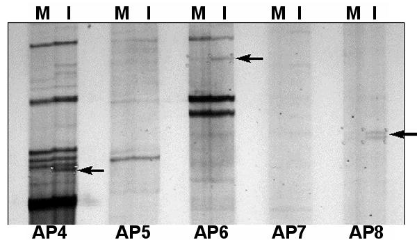
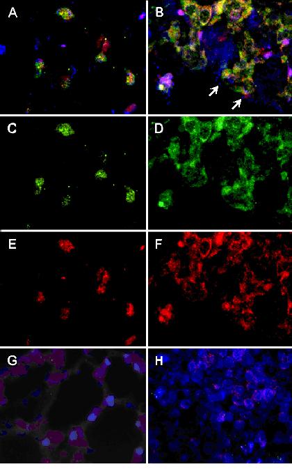
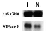
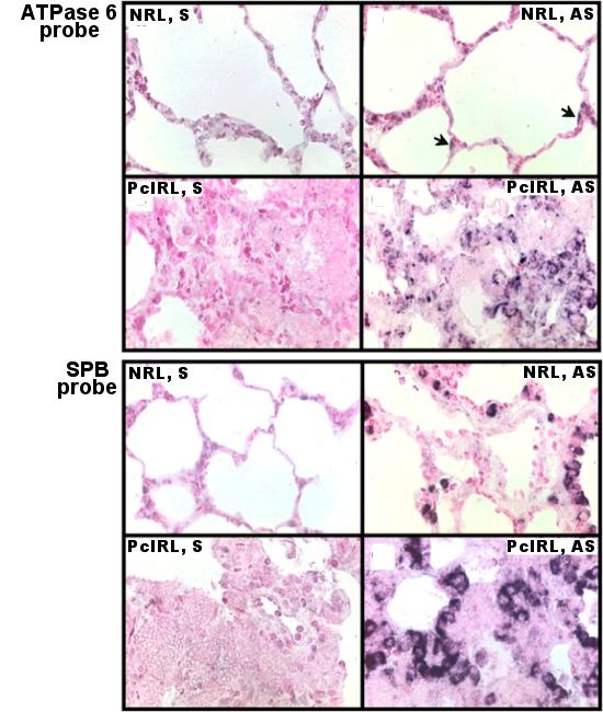
The technique of mRNA differential display was used to detect genes whose expression may be affected by P. carinii infection. The nucleotide sequence of one differentially displayed DNA fragment was found to be identical to that of the rat mitochondrial ATPase 6 gene, which is a subunit of the F0F1-ATP synthase complex. A four-fold increase in expression of this gene was verified by Northern blot analysis of total RNA extracted from P. carinii-infected rat lung versus that from mock-infected rat lung. Localization of the cells containing ATPase 6 mRNA was accomplished by in situ hybridization. In sections of non-infected rat lung, these cells were found lining the distal parts of the respiratory tree and in apical areas of the alveoli. Histological location of these cells suggested that they were Clara cells and type II pneumocytes. This hypothesis was confirmed by co-localizing the mRNAs for ATPase 6 and surfactant protein B (SP-B) to the same cells by two-color fluorescent in situ hybridization.
The ATPase 6 gene is over expressed during P. carinii infection, and type II pneumocytes and Clara cells are the cell types responsible for this over-expression.
卡氏肺孢子虫可导致免疫功能低下患者发生肺炎,其发病率和死亡率都很高,但该病原体与宿主细胞之间的相互作用尚未完全明确。本研究的目的是在分子水平上研究宿主细胞对卡氏肺孢子虫感染的反应。
采用mRNA差异显示技术检测可能受卡氏肺孢子虫感染影响的基因表达。发现一个差异显示DNA片段的核苷酸序列与大鼠线粒体ATP酶6基因相同,该基因是F0F1 - ATP合酶复合体的一个亚基。通过对从感染卡氏肺孢子虫的大鼠肺组织和未感染的对照大鼠肺组织中提取的总RNA进行Northern印迹分析,证实该基因的表达增加了四倍。通过原位杂交确定了含有ATP酶6 mRNA的细胞的定位。在未感染大鼠肺组织切片中,这些细胞位于呼吸树的远端部分和肺泡的顶端区域。这些细胞的组织学定位表明它们是克拉拉细胞和II型肺泡上皮细胞。通过双色荧光原位杂交将ATP酶6和表面活性蛋白B(SP - B)的mRNA共定位到同一细胞,证实了这一假设。
在卡氏肺孢子虫感染期间,ATP酶6基因过度表达,II型肺泡上皮细胞和克拉拉细胞是导致这种过度表达的细胞类型。