Gerber S A, Moran J P, Frelinger J G, Frelinger J A, Fenton B M, Lord E M
Department of Microbiology & Immunology, University of Rochester Medical Center, Rochester, NY 14642, USA.
Br J Cancer. 2003 May 6;88(9):1453-61. doi: 10.1038/sj.bjc.6600907.
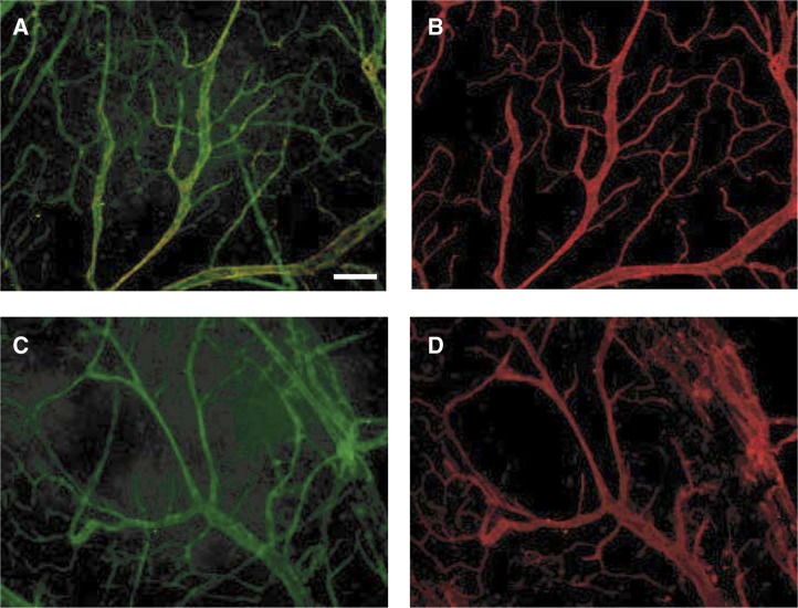
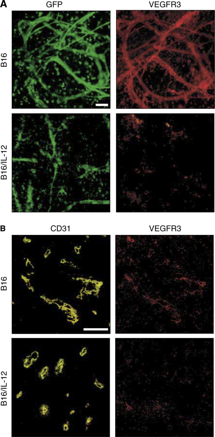
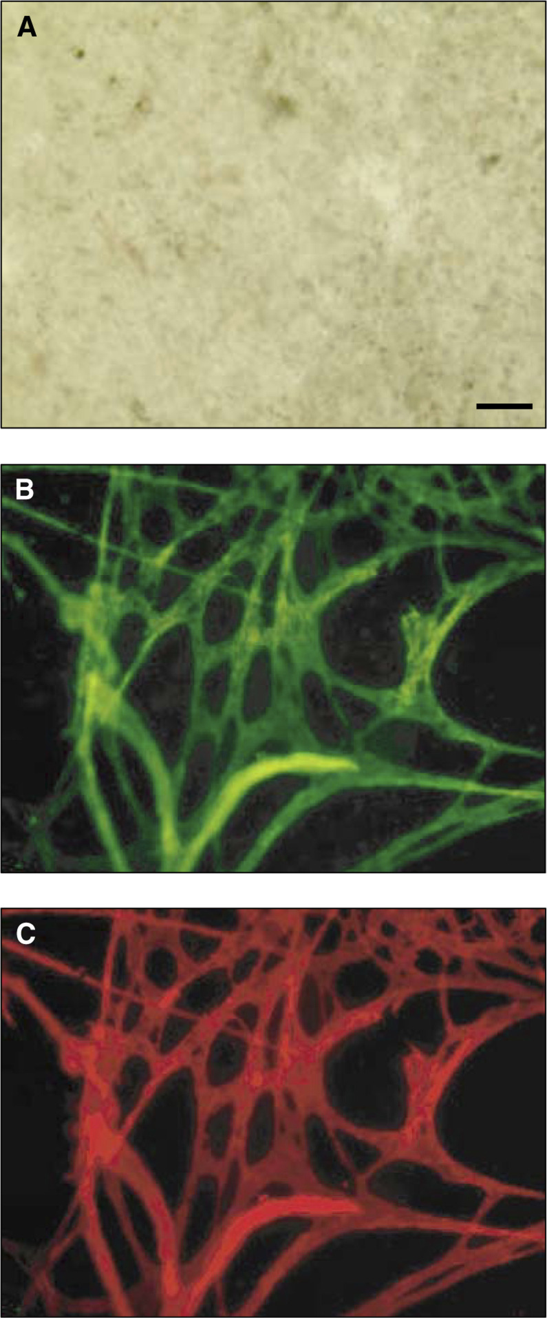
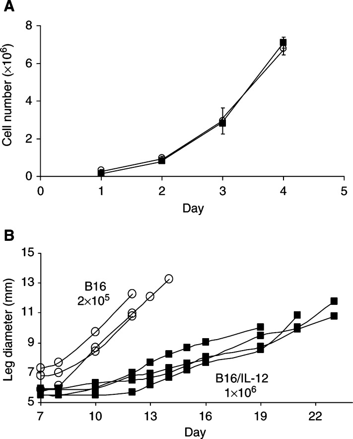
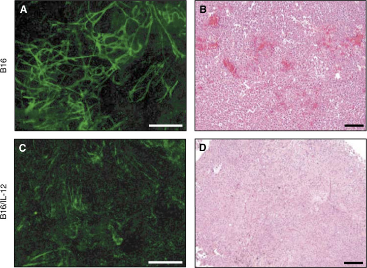
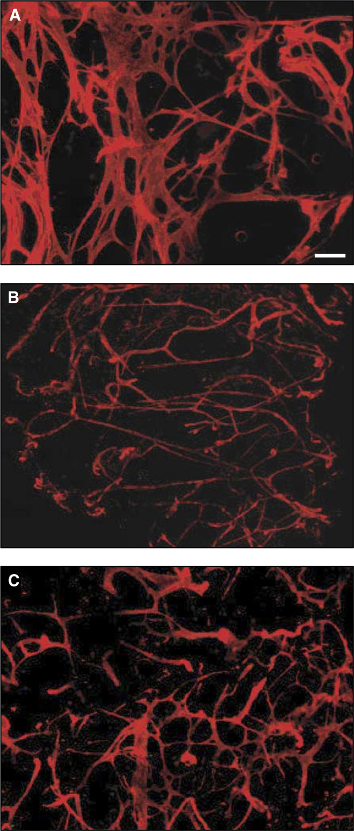
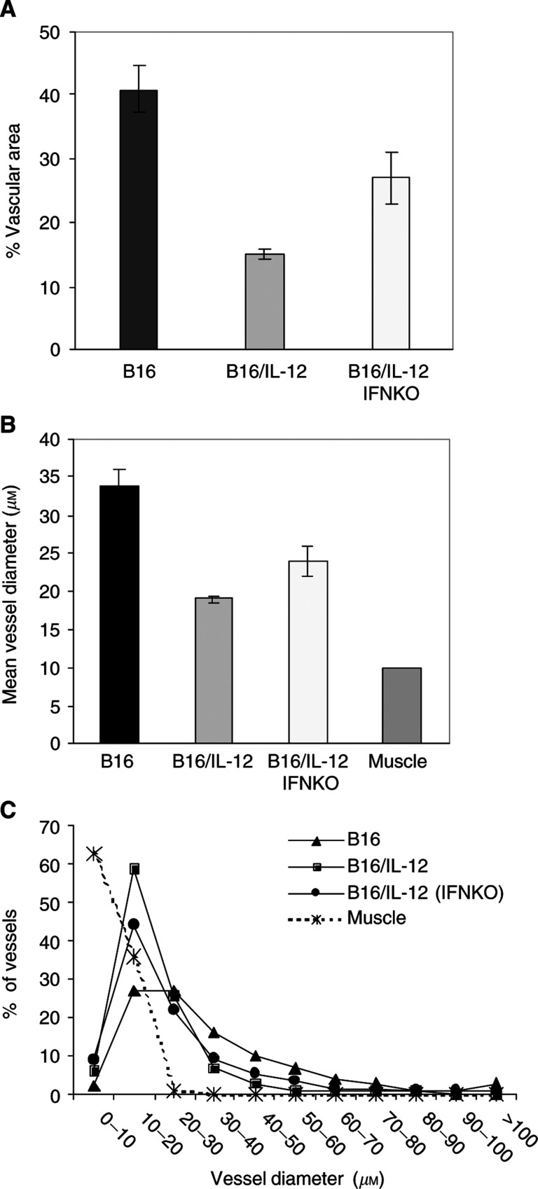
New blood vessel formation within tumours is a critical feature for tumour growth. A major limitation in understanding this complex process has been the inability to visualise and analyse vessel formation. Here, we report on the development of a whole-tissue mount technique that allows visualisation of vessel structure. Mice expressing green fluorescent protein (GFP) made it possible to easily see GFP(+) vessels within non-GFP-expressing B16 melanoma tumours. The small fragments of tumour used in this technique were also effectively stained with fluorescent probe-conjugated antibodies, allowing characterisation of the vessels based on surface marker phenotype. The vessels within tumour tissue were much more irregular and tortuous compared to those within surrounding normal muscle. B16 tumours stably transfected with the genes for IL-12 were used to assess the effects of this cytokine on tumour growth and vessel formation. The IL-12-expressing tumours grew more slowly and had much smaller blood vessels than the large, webbed vessels characteristic of the parental tumours, effects that were dependent on interferon gamma (IFN-gamma). Vessels in the parental tumours were found to express VEGFR-3, the receptor for VEGF-C and VEGF-D. Expression of this receptor by the endothelial cells of the blood vessels was lost in the cytokine expressing tumours, thus suggesting a mechanism for the antiangiogenic effects of IL-12. The combination of the whole mount technique and the GFP transgenic mice provides a powerful method for visualising tumour vasculature and characterising the effects of agents such as cytokines.
肿瘤内新血管形成是肿瘤生长的关键特征。理解这一复杂过程的一个主要限制是无法可视化和分析血管形成。在此,我们报告一种全组织贴片技术的开发,该技术可实现血管结构的可视化。表达绿色荧光蛋白(GFP)的小鼠使得在不表达GFP的B16黑色素瘤肿瘤内轻松看到GFP(+)血管成为可能。该技术中使用的肿瘤小片段也能用荧光探针偶联抗体有效染色,从而基于表面标志物表型对血管进行表征。与周围正常肌肉中的血管相比,肿瘤组织内的血管更加不规则且蜿蜒曲折。用IL-12基因稳定转染的B16肿瘤用于评估这种细胞因子对肿瘤生长和血管形成的影响。表达IL-12的肿瘤生长更缓慢,且血管比亲本肿瘤特有的大的、网状血管小得多,这些效应依赖于干扰素γ(IFN-γ)。发现亲本肿瘤中的血管表达VEGFR-3,即VEGF-C和VEGF-D的受体。在表达细胞因子的肿瘤中,血管内皮细胞失去了该受体的表达,因此提示了IL-12抗血管生成作用的一种机制。全贴片技术和GFP转基因小鼠的结合为可视化肿瘤脉管系统以及表征细胞因子等试剂的作用提供了一种强大的方法。