Devoto Paola, Flore Giovanna, Saba Pierluigi, Fà Mauro, Gessa Gian Luigi
Department of Neuroscience B.B. Brodie University of Cagliari, Italy.
BMC Neurosci. 2005 May 2;6:31. doi: 10.1186/1471-2202-6-31.
Previous studies by our group suggest that extracellular dopamine (DA) and noradrenaline (NA) may be co-released from noradrenergic nerve terminals in the cerebral cortex. We recently demonstrated that the concomitant release of DA and NA could be elicited in the cerebral cortex by electrical stimulation of the locus coeruleus (LC). This study analyses the effect of both single train and repeated electrical stimulation of LC on NA and DA release in the medial prefrontal cortex (mPFC), occipital cortex (Occ), and caudate nucleus. To rule out possible stressful effects of electrical stimulation, experiments were performed on chloral hydrate anaesthetised rats.
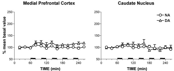
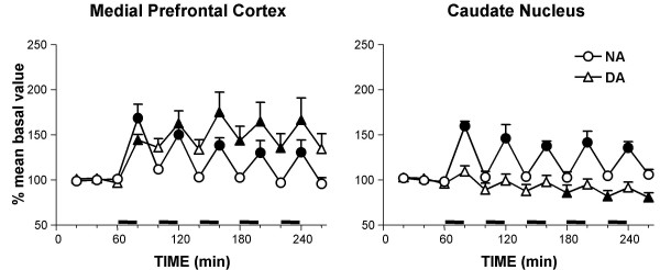
Twenty min electrical stimulation of the LC, with burst type pattern of pulses, increased NA and DA both in the mPFC and in the Occ. NA in both cortices and DA in the mPFC returned to baseline within 20 min after the end of the stimulation period, while DA in the Occ reached a maximum increase during 20 min post-stimulation and remained higher than baseline values at 220 min post-stimulation. Local perfusion with tetrodotoxin (TTX, 10 microM) markedly reduced baseline NA and DA in the mPFC and Occ and totally suppressed the effect of electrical stimulation in both areas. A sequence of five 20 min stimulations at 20 min intervals were delivered to the LC. Each stimulus increased NA to the same extent and duration as the first stimulus, whereas DA remained elevated at the time next stimulus was delivered, so that baseline DA progressively increased in the mPFC and Occ to reach about 130 and 200% the initial level, respectively. In the presence of the NA transport (NAT) blocker desipramine (DMI, 100 microM), multiple LC stimulation still increased extracellular NA and DA levels. Electrical stimulation of the LC increased NA levels in the homolateral caudate nucleus, but failed to modify DA level.
The results confirm and extend that LC stimulation induces a concomitant release of DA and NA in the mPFC and Occ. The different time-course of LC-induced elevation of DA and NA suggests that their co-release may be differentially controlled.
我们团队之前的研究表明,细胞外多巴胺(DA)和去甲肾上腺素(NA)可能从大脑皮质的去甲肾上腺素能神经末梢共同释放。我们最近证明,通过电刺激蓝斑(LC)可在大脑皮质引发DA和NA的伴随释放。本研究分析了对LC进行单次串刺激和重复电刺激对内侧前额叶皮质(mPFC)、枕叶皮质(Occ)和尾状核中NA和DA释放的影响。为排除电刺激可能产生的应激效应,实验在水合氯醛麻醉的大鼠身上进行。
对LC进行20分钟的脉冲串模式电刺激,可使mPFC和Occ中的NA和DA均增加。刺激期结束后20分钟内,两个皮质中的NA以及mPFC中的DA均恢复至基线水平,而Occ中的DA在刺激后20分钟达到最大增加值,并在刺激后220分钟仍高于基线值。用河豚毒素(TTX,10微摩尔)局部灌注可显著降低mPFC和Occ中的基线NA和DA,并完全抑制两个区域的电刺激效应。以20分钟的间隔对LC进行一系列五次20分钟的刺激。每次刺激使NA增加的程度和持续时间与第一次刺激相同,而在下一次刺激施加时DA仍保持升高,因此mPFC和Occ中的基线DA逐渐增加,分别达到初始水平的约130%和200%。在存在去甲肾上腺素转运体(NAT)阻断剂地昔帕明(DMI,100微摩尔)的情况下,多次LC刺激仍可增加细胞外NA和DA水平。电刺激LC可使同侧尾状核中的NA水平升高,但未能改变DA水平。
结果证实并扩展了LC刺激可在mPFC和Occ中诱导DA和NA伴随释放的观点。LC诱导的DA和NA升高的不同时间进程表明,它们的共同释放可能受到不同的调控。