Kurtti Timothy J, Simser Jason A, Baldridge Gerald D, Palmer Ann T, Munderloh Ulrike G
Department of Entomology, University of Minnesota, St. Paul, MN 55108, USA.
J Invertebr Pathol. 2005 Nov;90(3):177-86. doi: 10.1016/j.jip.2005.09.001. Epub 2005 Nov 9.
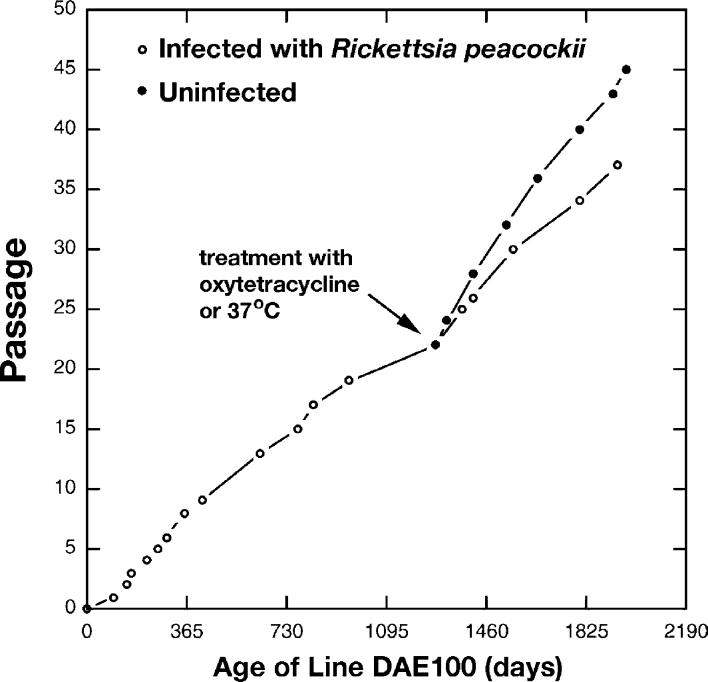
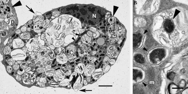
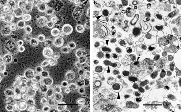
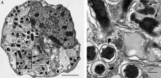
Rickettsia peacockii, a spotted fever group rickettsia, is a transovarially transmitted endosymbiont of Rocky Mountain wood ticks, Dermacentor andersoni. This rickettsia, formerly known as the East Side Agent and restricted to female ticks, was detected in a chronically infected embryonic cell line, DAE100, from D. andersoni. We examined infectivity, ability to induce cytopathic effect (CPE) and host cell specificity of R. peacockii using cultured arthropod and mammalian cells. Aposymbiotic DAE100 cells were obtained using oxytetracycline or incubation at 37 degrees C. Uninfected DAE100 sublines grew faster than the parent line, indicating R. peacockii regulation of host cell growth. Nevertheless, DAE100 cellular defenses exerted partial control over R. peacockii growth. Rickettsiae existed free in the cytosol of DAE100 cells or within autophagolysosomes. Exocytosed rickettsiae accumulated in the medium and were occasionally contained within host membranes. R. peacockii multiplied in other cell lines from the hard ticks D. andersoni, Dermacentor albipictus, Ixodes scapularis, and Ixodes ricinus; the soft tick Carios capensis; and the lepidopteran Trichoplusia ni. Lines from the tick Amblyomma americanum, the mosquito Aedes albopictus, and two mammalian cell lines were non-permissive to R. peacockii. High cell densities facilitated rickettsial spread within permissive cell cultures, and an inoculum of one infected to nine uninfected cells resulted in the greatest yield of infected tick cells. Cell-free R. peacockii also were infectious for tick cells and centrifugation onto cell layers enhanced infectivity approximately 100-fold. The ability of R. peacockii to cause mild CPE suggests that its pathogenicity is not completely muted. An analysis of R. peacockii-cell interactions in comparison to pathogenic rickettsiae will provide insights into host cell colonization mechanisms.
孔雀立克次氏体是斑点热群立克次氏体,是落基山木蜱(安氏革蜱)经卵传播的内共生菌。这种立克次氏体以前被称为东区病原体,且仅存在于雌性蜱中,在一株来自安氏革蜱的慢性感染胚胎细胞系DAE100中被检测到。我们使用培养的节肢动物和哺乳动物细胞,研究了孔雀立克次氏体的感染性、诱导细胞病变效应(CPE)的能力以及宿主细胞特异性。使用土霉素或在37℃孵育获得无共生体的DAE100细胞。未感染的DAE100亚系比亲代细胞系生长得更快,这表明孔雀立克次氏体对宿主细胞生长有调节作用。然而,DAE100细胞的防御机制对孔雀立克次氏体的生长有部分控制作用。立克次氏体在DAE100细胞的胞质溶胶中自由存在或存在于自噬溶酶体内。胞吐的立克次氏体积聚在培养基中,偶尔被宿主膜包裹。孔雀立克次氏体在来自硬蜱安氏革蜱、白纹革蜱、肩突硬蜱和蓖麻硬蜱;软蜱卡氏钝缘蜱;以及鳞翅目粉纹夜蛾的其他细胞系中增殖。来自美洲钝眼蜱、白纹伊蚊的细胞系以及两种哺乳动物细胞系对孔雀立克次氏体不敏感。高细胞密度促进了立克次氏体在允许的细胞培养物中的传播,一份感染细胞与九份未感染细胞的接种物可使感染的蜱细胞产量最高。无细胞的孔雀立克次氏体对蜱细胞也具有感染性,离心接种到细胞层上可使感染性提高约100倍。孔雀立克次氏体引起轻度CPE的能力表明其致病性并未完全消失。将孔雀立克次氏体与致病性立克次氏体的细胞相互作用进行分析,将有助于深入了解宿主细胞定植机制。