Chen Gang, Khalil Nasreen
Division of Respiratory Medicine, Department of Medicine, The University of British Columbia, The Vancouver Coastal Health Research Institute, Vancouver, BC V6H 3Z6, Canada.
Respir Res. 2006 Jan 3;7(1):2. doi: 10.1186/1465-9921-7-2.
Airway remodeling in asthma is the result of increased expression of connective tissue proteins, airway smooth muscle cell (ASMC) hyperplasia and hypertrophy. TGF-beta1 has been found to increase ASMC proliferation. The activation of mitogen-activated protein kinases (MAPKs), p38, ERK, and JNK, is critical to the signal transduction associated with cell proliferation. In the present study, we determined the role of phosphorylated MAPKs in TGF-beta1 induced ASMC proliferation.
Confluent and growth-arrested bovine ASMCs were treated with TGF-beta1. Proliferation was measured by [3H]-thymidine incorporation and cell counting. Expressions of phosphorylated p38, ERK1/2, and JNK were determined by Western analysis.
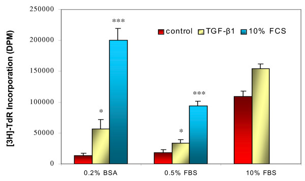
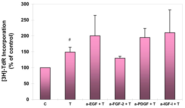
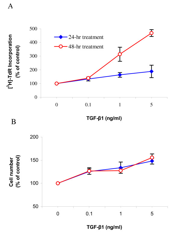
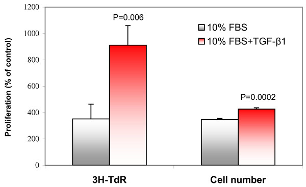
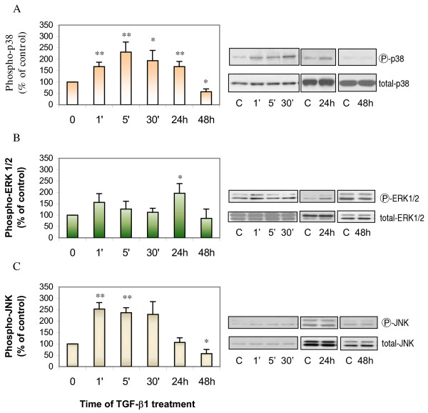
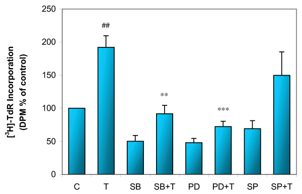
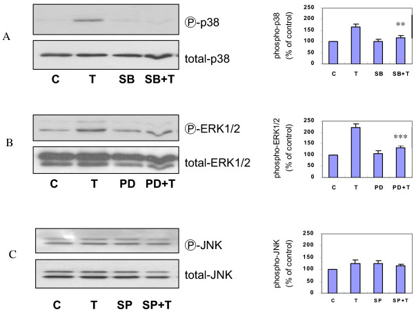
In a concentration-dependent manner, TGF-beta1 increased [3H]-thymidine incorporation and cell number of ASMCs. TGF-beta1 also enhanced serum-induced ASMC proliferation. Although ASMCs cultured with TGF-beta1 had a significant increase in phosphorylated p38, ERK1/2, and JNK, the maximal phosphorylation of each MAPK had a varied onset after incubation with TGF-beta1. TGF-beta1 induced DNA synthesis was inhibited by SB 203580 or PD 98059, selective inhibitors of p38 and MAP kinase kinase (MEK), respectively. Antibodies against EGF, FGF-2, IGF-I, and PDGF did not inhibit the TGF-beta1 induced DNA synthesis.
Our data indicate that ASMCs proliferate in response to TGF-beta1, which is mediated by phosphorylation of p38 and ERK1/2. These findings suggest that TGF-beta1 which is expressed in airways of asthmatics may contribute to irreversible airway remodeling by enhancing ASMC proliferation.
哮喘中的气道重塑是结缔组织蛋白表达增加、气道平滑肌细胞(ASMC)增生和肥大的结果。已发现转化生长因子-β1(TGF-β1)可增加ASMC增殖。丝裂原活化蛋白激酶(MAPK)p38、ERK和JNK的激活对于与细胞增殖相关的信号转导至关重要。在本研究中,我们确定了磷酸化MAPK在TGF-β1诱导的ASMC增殖中的作用。
用TGF-β1处理汇合且生长停滞的牛ASMC。通过[3H] -胸苷掺入和细胞计数测量增殖。通过蛋白质印迹分析确定磷酸化p38、ERK1/2和JNK的表达。
TGF-β1以浓度依赖的方式增加ASMC的[3H] -胸苷掺入和细胞数量。TGF-β1还增强了血清诱导的ASMC增殖。尽管用TGF-β1培养的ASMC中磷酸化p38、ERK1/2和JNK有显著增加,但与TGF-β1孵育后,每种MAPK的最大磷酸化出现时间不同。分别用p38和丝裂原活化蛋白激酶激酶(MEK)的选择性抑制剂SB 203580或PD 98059抑制TGF-β1诱导的DNA合成。针对表皮生长因子(EGF)、成纤维细胞生长因子-2(FGF-2)、胰岛素样生长因子-I(IGF-I)和血小板衍生生长因子(PDGF)的抗体不抑制TGF-β1诱导的DNA合成。
我们的数据表明,ASMC对TGF-β1作出反应而增殖,这是由p38和ERK1/2的磷酸化介导的。这些发现表明,哮喘患者气道中表达的TGF-β1可能通过增强ASMC增殖而导致不可逆的气道重塑。