Zannoni Augusta, Bernardini Chiara, Rada Tommaso, Ribeiro Luciana A, Forni Monica, Bacci Maria L
Department of Veterinary Morphophysiology and Animal Production, DIMORFIPA, Ozzano Emilia, University of Bologna, Italy.
Reprod Biol Endocrinol. 2007 Jul 20;5:31. doi: 10.1186/1477-7827-5-31.
The corpus luteum (CL) is a transient endocrine gland and prostaglandin F2-alpha is considered to be the principal luteolysin in pigs. In this species, the in vivo administration of prostaglandin F2-alpha induces apoptosis in large vessels as early as 6 hours after administration. The presence of the prostaglandin F2-alpha receptor (FPr) on the microvascular endothelial cells (pCL-MVECs) of the porcine corpus luteum has not yet been defined. The aim of the study was to assess FPr expression in pCL-MVECs in the early and mid-luteal phases (EL-p, ML-p), and during pregnancy (P-p). Moreover, the effectiveness of prostaglandin F2-alpha treatment in inducing pCL-MVEC apoptosis was tested.
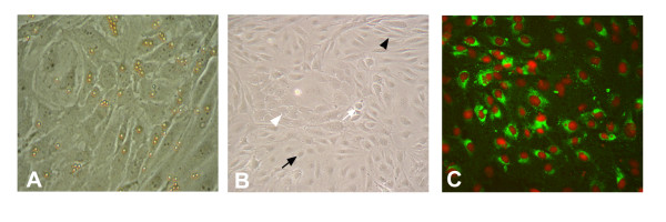
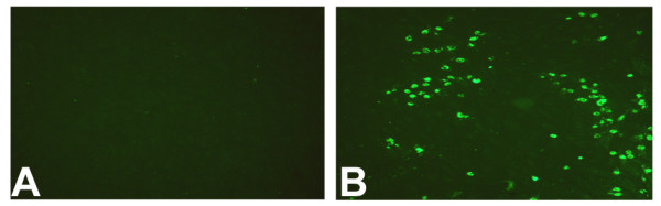
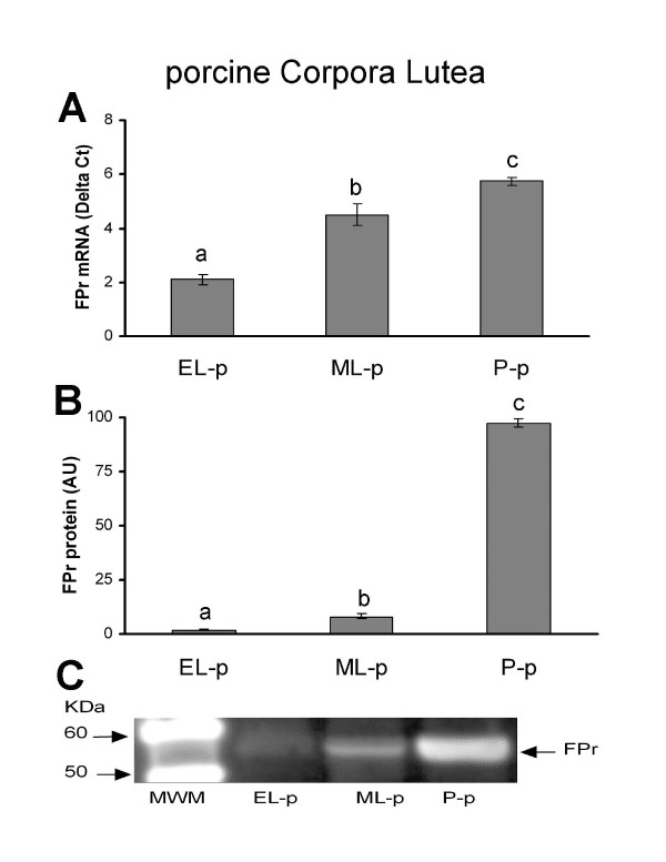
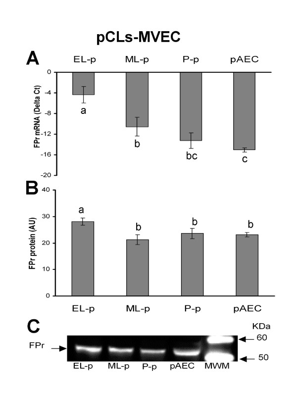
Porcine CLs were collected in the EL and ML phases and during P-p. All CLs from each animal were minced together and the homogenates underwent enzymatic digestion. The pCL-MVECs were then positively selected by an immunomagnetic separation protocol using Dynabeads coated with anti-CD31 monoclonal antibody and seeded in flasks in the presence of EGM 2-MV (Microvascular Endothelial Cell Medium-2). After 4 days of culture, the cells underwent additional immunomagnetic selection and were seeded in flasks until the confluent stage.PCR Real time, western blot and immunodetection assays were utilized to assess the presence of FPr on pCL-MVEC primary cultures. Furthermore, the influence of culture time (freshly isolated, cultured overnight and at confluence) and hormonal treatment (P4 and E2) on FPr expression in pCL-MVECs was also investigated. Apoptosis was detected by TUNEL assay of pCL-MVECs exposed to prostaglandin F2-alpha.
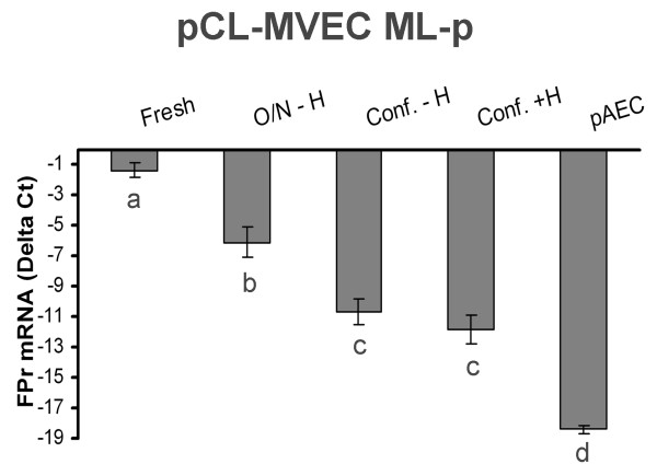
We obtained primary cultures of pCL-MVECs from all animals. FPr mRNA and protein levels showed the highest value (ANOVA) in CL-MVECs derived from the early-luteal phase. Moreover, freshly isolated MVECs showed a higher FPr mRNA value than those cultured overnight and confluent cells (ANOVA). prostaglandin F2-alpha treatment failed to induce an apoptotic response in all the pCL-MVEC cultures.
Our data showing the presence of FPr on MVECs and the inability of prostaglandin F2-alpha to evoke an in vitro apoptotic response suggest that other molecules or mechanisms must be considered in order to explain the in vivo direct pro-apoptotic effect of prostaglandin F2-alpha at the endothelial level.
黄体(CL)是一种临时性内分泌腺,前列腺素F2α被认为是猪体内主要的溶黄体素。在该物种中,体内注射前列腺素F2α最早在注射后6小时就会诱导大血管中的细胞凋亡。猪黄体微血管内皮细胞(pCL-MVECs)上前列腺素F2α受体(FPr)的存在尚未明确。本研究的目的是评估FPr在黄体早期和中期(EL-p、ML-p)以及妊娠期(P-p)的pCL-MVECs中的表达情况。此外,还测试了前列腺素F2α处理诱导pCL-MVECs凋亡的有效性。
在黄体早期和中期以及妊娠期收集猪的黄体。将每只动物的所有黄体切碎,匀浆进行酶消化。然后使用包被有抗CD31单克隆抗体的磁珠通过免疫磁珠分离方案对pCL-MVECs进行阳性选择,并在含有EGM 2-MV(微血管内皮细胞培养基-2)的培养瓶中接种。培养4天后,对细胞进行额外的免疫磁珠选择,并接种到培养瓶中直至汇合阶段。利用实时PCR、蛋白质印迹和免疫检测分析评估pCL-MVEC原代培养物中FPr的存在情况。此外,还研究了培养时间(新鲜分离、过夜培养和汇合)和激素处理(P4和E2)对pCL-MVECs中FPr表达的影响。通过对暴露于前列腺素F2α的pCL-MVECs进行TUNEL分析检测细胞凋亡。
我们从所有动物中获得了pCL-MVECs的原代培养物。FPr mRNA和蛋白水平在来自黄体早期的CL-MVECs中显示出最高值(方差分析)。此外,新鲜分离的MVECs显示出比过夜培养和汇合细胞更高的FPr mRNA值(方差分析)。前列腺素F2α处理未能在所有pCL-MVEC培养物中诱导凋亡反应。
我们的数据表明MVECs上存在FPr,且前列腺素F2α无法在体外引发凋亡反应,这表明为了解释前列腺素F2α在体内对内皮细胞水平的直接促凋亡作用,必须考虑其他分子或机制。