Radley Jason J, Rocher Anne B, Rodriguez Alfredo, Ehlenberger Douglas B, Dammann Mark, McEwen Bruce S, Morrison John H, Wearne Susan L, Hof Patrick R
Laboratory of Neuronal Structure and Function, Salk Institute for Biological Studies, La Jolla, California 92037, USA.
J Comp Neurol. 2008 Mar 1;507(1):1141-50. doi: 10.1002/cne.21588.
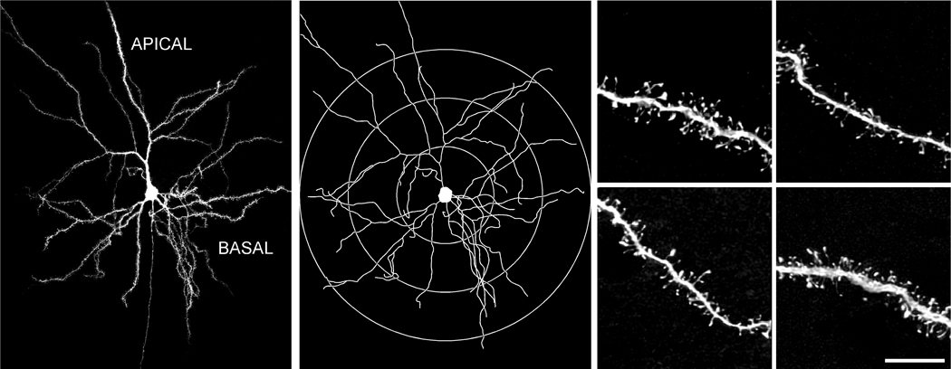
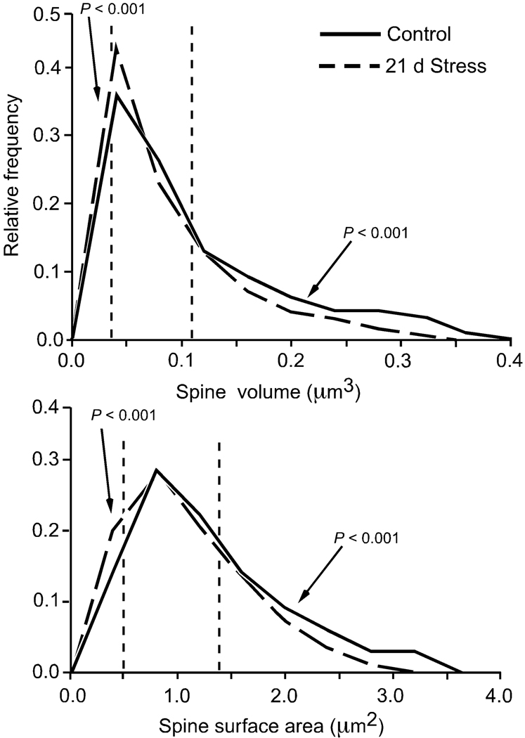
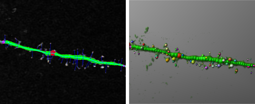
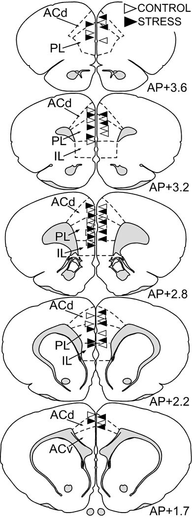
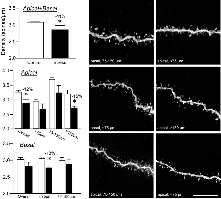
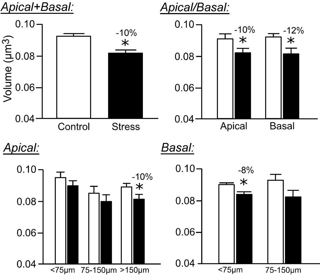
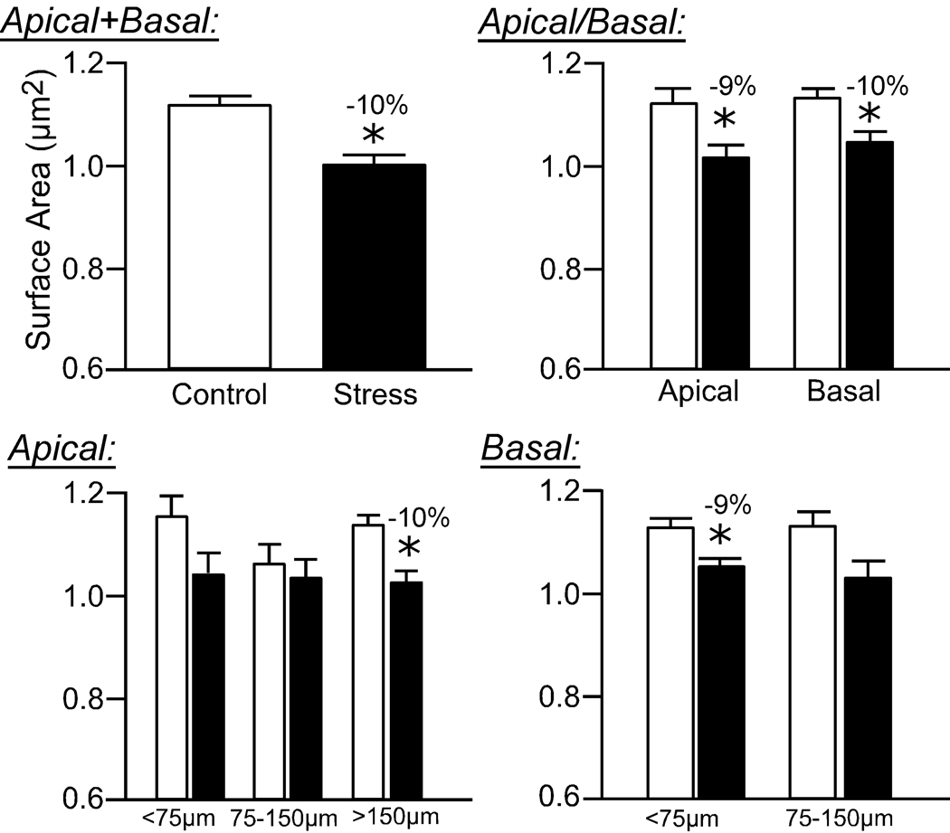
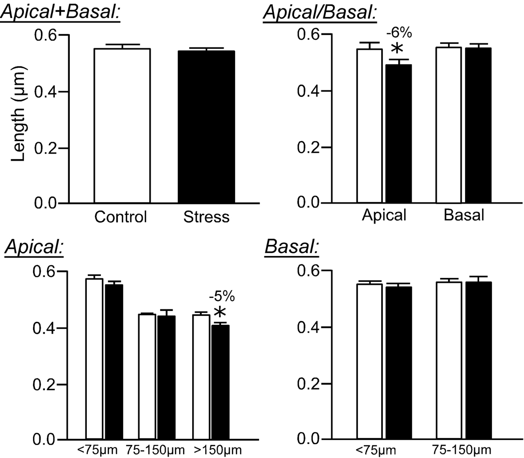
Anatomical alterations in the medial prefrontal cortex (mPFC) are associated with hypothalamopituitary adrenal (HPA) axis dysregulation, altered stress hormone levels, and psychiatric symptoms of stress-related mental illnesses. Functional imaging studies reveal impairment and shrinkage of the mPFC in such conditions, and these findings are paralleled by experimental studies showing dendritic retraction and spine loss following repeated stress in rodents. Here we extend this characterization to how repeated stress affects dendritic spine morphology in mPFC through the utilization of an automated approach that rapidly digitizes, reconstructs three dimensionally, and calculates geometric features of neurons. Rats were perfused after being subjected to 3 weeks of daily restraint stress (6 hours/day), and intracellular injections of Lucifer Yellow were made in layer II/III pyramidal neurons in the dorsal mPFC. To reveal spines in all angles of orientation, deconvolved high-resolution confocal laser scanning microscopy image stacks of dendritic segments were reconstructed and analyzed for spine volume, surface area, and length using a Rayburst-based automated approach (8,091 and 8,987 spines for control and stress, respectively). We found that repeated stress results in an overall decrease in mean dendritic spine volume and surface area, which was most pronounced in the distal portion of apical dendritic fields. Moreover, we observed an overall shift in the population of spines, manifested by a reduction in large spines and an increase in small spines. These results suggest a failure of spines to mature and stabilize following repeated stress and are likely to have major repercussions on function, receptor expression, and synaptic efficacy.
内侧前额叶皮质(mPFC)的解剖学改变与下丘脑 - 垂体 - 肾上腺(HPA)轴功能失调、应激激素水平改变以及应激相关精神疾病的精神症状有关。功能成像研究显示,在这些情况下mPFC会出现损伤和萎缩,实验研究也证实了这一点,即啮齿动物反复应激后会出现树突回缩和棘突丢失。在此,我们通过一种自动化方法扩展了对反复应激如何影响mPFC中树突棘形态的描述,该方法可快速数字化、三维重建并计算神经元的几何特征。大鼠在接受为期3周的每日束缚应激(6小时/天)后进行灌注,然后向背侧mPFC的II/III层锥体神经元内注射荧光黄。为了从各个方向揭示棘突,对去卷积后的树突节段高分辨率共聚焦激光扫描显微镜图像堆栈进行重建,并使用基于Rayburst的自动化方法分析棘突的体积、表面积和长度(对照组和应激组分别有8091个和8987个棘突)。我们发现,反复应激导致平均树突棘体积和表面积总体减小,这在顶端树突场的远端最为明显。此外,我们观察到棘突群体发生了整体变化,表现为大棘突减少,小棘突增加。这些结果表明,反复应激后棘突未能成熟和稳定,这可能对功能、受体表达和突触效能产生重大影响。