Rostron Claire L, Farquhar Morag J, Latimer Mary P, Winn Philip
Life Sciences, The Open University, Walton Hall, Milton Keynes, MK7 6AA, UK.
BMC Neurosci. 2008 Jan 30;9:16. doi: 10.1186/1471-2202-9-16.
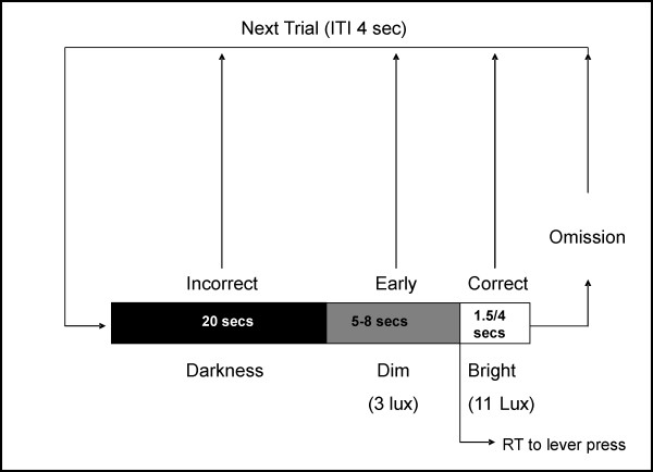
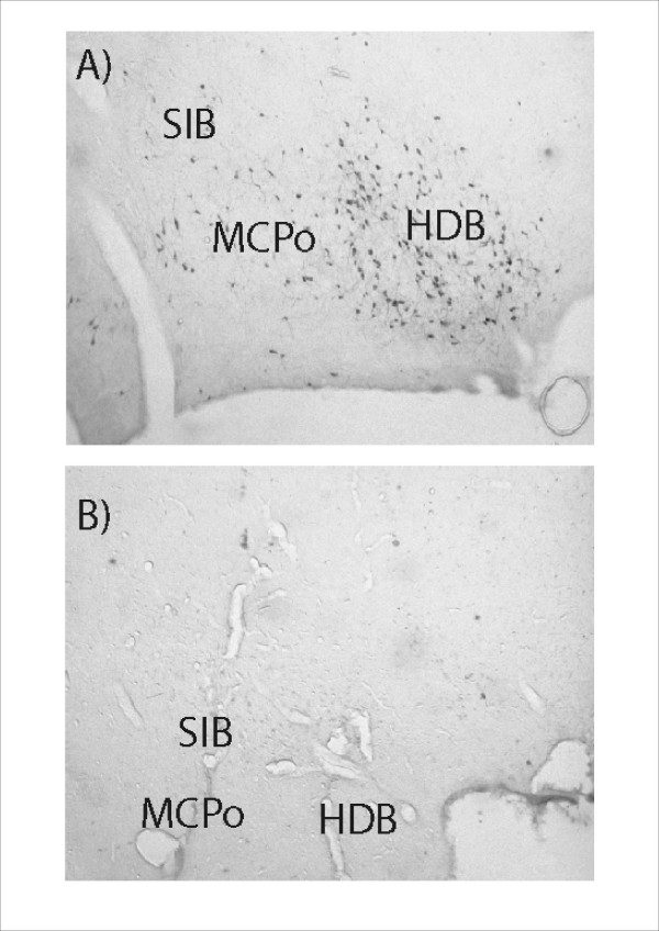
It is well established that nucleus basalis magnocellularis (NbM) lesions impair performance on tests of sustained attention. Previous work from this laboratory has also demonstrated that pedunculopontine tegmental nucleus (PPTg) lesioned rats make more omissions on a test of sustained attention, suggesting that it might also play a role in mediating this function. However, the results of the PPTg study were open to alternative interpretation. We aimed to resolve this by conducting a detailed analysis of the effects of damage to each brain region in the same sustained attention task used in our previous work. Rats were trained in the task before surgery and post-surgical testing examined performance in response to unpredictable light signals of 1500 ms and 4000 ms duration. Data for PPTg lesioned rats were compared to control rats, and rats with 192 IgG saporin infusions centred on the NbM. In addition to operant data, video data of rats' performance during the task were also analysed.
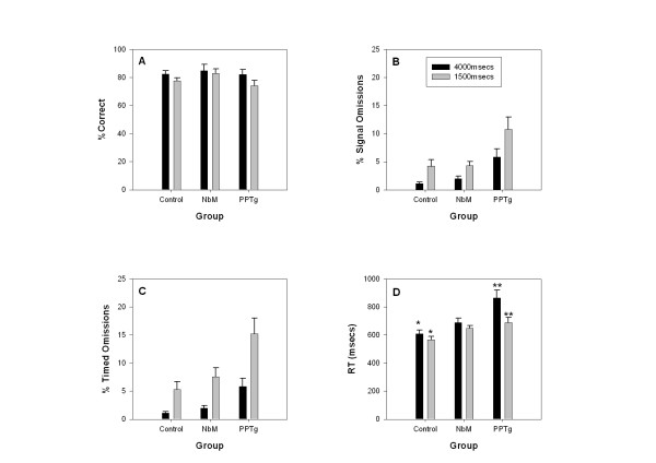
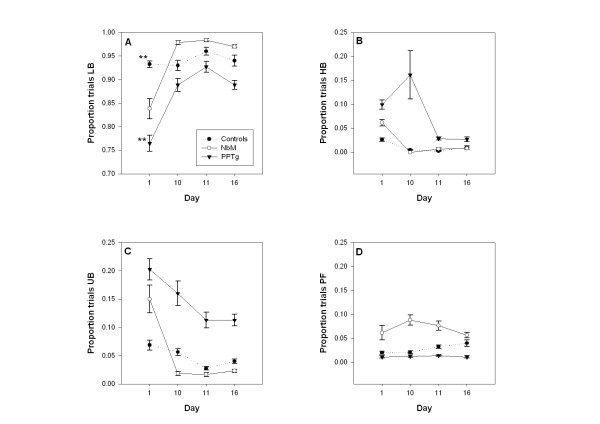
Both lesion groups omitted trials relative to controls but the effect was milder and transient in NbM rats. The number of omitted trials decreased in all groups when tested using the 4000 ms signal compared to the 1500 ms signal. This confirmed previous findings for PPTg lesioned rats. Detailed analysis revealed that the increase in omissions in PPTg rats was not a consequence of motor impairment. The video data (taken on selected days) showed reduced lever orientation in PPTg lesioned rats, coupled with an increase in unconditioned behaviours such as rearing and sniffing. In contrast NbM rats showed evidence of inadequate lever pressing.
The question addressed here is whether the PPTg and NbM both have a role in sustained attention. Rats bearing lesions of either structure showed deficits in the test used. However, we conclude that the most parsimonious explanation for the deficit observed in PPTg rats is inadequate response organization, rather than impairment in sustained attention. Furthermore the impairment observed in NbM lesioned rats included lever pressing difficulties in addition to impaired sustained attention. Unfortunately we could not link these deficits directly to cholinergic neuronal loss.
已有充分证据表明,基底大细胞核(NbM)损伤会损害持续注意力测试中的表现。本实验室先前的研究还表明,脚桥被盖核(PPTg)损伤的大鼠在持续注意力测试中出现更多遗漏情况,这表明它可能也在介导该功能中发挥作用。然而,PPTg研究的结果存在其他解释的可能性。我们旨在通过对与先前工作相同的持续注意力任务中每个脑区损伤的影响进行详细分析来解决这一问题。大鼠在手术前接受该任务的训练,术后测试检查其对持续时间为1500毫秒和4000毫秒的不可预测光信号的反应表现。将PPTg损伤大鼠的数据与对照大鼠以及以NbM为中心注射192IgG皂草素的大鼠的数据进行比较。除了操作性数据外,还分析了大鼠在任务期间表现的视频数据。
与对照组相比,两个损伤组都有遗漏试验的情况,但在NbM大鼠中这种影响较轻微且短暂。与使用1500毫秒信号进行测试相比,当使用4000毫秒信号进行测试时,所有组遗漏试验的数量均减少。这证实了先前关于PPTg损伤大鼠的研究结果。详细分析表明,PPTg大鼠遗漏情况的增加并非运动障碍的结果。视频数据(在选定的日子采集)显示,PPTg损伤大鼠的杠杆定向减少,同时诸如竖毛和嗅探等非条件行为增加。相比之下,NbM大鼠表现出杠杆按压不足的迹象。
这里探讨的问题是PPTg和NbM是否都在持续注意力中起作用。患有这两种结构损伤的大鼠在所用测试中均表现出缺陷。然而,我们得出的结论是,对PPTg大鼠中观察到的缺陷最简洁的解释是反应组织不足,而非持续注意力受损。此外,在NbM损伤大鼠中观察到的损伤除了持续注意力受损外,还包括杠杆按压困难。不幸的是,我们无法将这些缺陷直接与胆碱能神经元丧失联系起来。