Dou Ying, Andersson-Lendahl Monika, Arner Anders
Department of Physiology and Pharmacology, Karolinska Institutet, Stockholm, Sweden.
J Gen Physiol. 2008 May;131(5):445-53. doi: 10.1085/jgp.200809982.
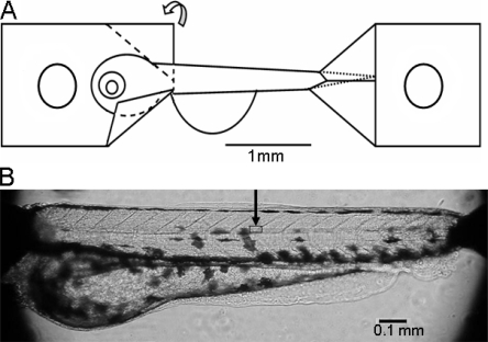
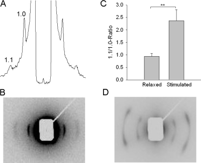
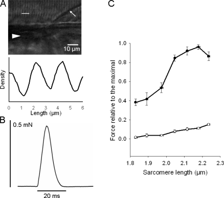
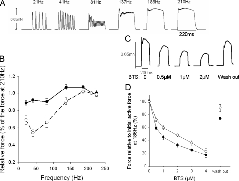
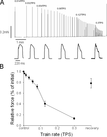
Zebrafish muscles were examined at an early developmental stage (larvae 5-7 d). Using aluminum clips, preparations (approximately 1.5 mm length, 150 microm diameter) were mounted for force registration and small angle x-ray diffraction. Sarcomeres were oriented mainly in parallel with the preparation long axis. Electrical stimulation elicited fast and reproducible single twitch contractions. Length-force relations showed an optimal sarcomere length of 2.15 microm. X-ray diffraction revealed clear equatorial 1.1/1.0 reflections, showing that myofilaments are predominantly arranged along the preparation long axis. In contrast, reflections from older (2 mo) zebrafish showed two main filament orientations each at an approximately 25 degrees angle relative to the preparation long axis. Electrical stimulation of larvae muscles increased the 1.1/1.0 intensity ratio, reflecting mass transfer to thin filaments during contraction. The apparent lattice volume was 3.42 x 10(-3) microm(3), which is smaller than that of mammalian striated muscle and more similar to that of frog muscles. The relation between force and stimulation frequency showed fusion of responses at a comparatively high frequency (approximately 186 Hz), reflecting a fast muscle phenotype. Inhibition of fast myosin with N-benzyl-p-toluene sulphonamide (BTS) showed that the later phase of the tetanus was less affected than the initial peak. This suggests that, although the main contractile phenotype is fast, slow twitch fibers can contribute to sustained contraction. A fatigue stimulation protocol with repeated 220 ms/186 Hz tetani showed that tetanic force decreased to 50% at a train rate of 0.1 s(-1). In conclusion, zebrafish larvae muscles can be examined in vitro using mechanical and x-ray methods. The muscles and myofilaments are mainly orientated in parallel with the larvae long axis and exhibit a significant fast contractile component. Sustained contractions can also involve a small contribution from slower muscle types.
在早期发育阶段(5 - 7日龄幼虫)对斑马鱼肌肉进行了检查。使用铝夹固定样本(长度约1.5毫米,直径150微米)用于力量记录和小角度X射线衍射。肌节主要与样本长轴平行排列。电刺激引发快速且可重复的单收缩。长度 - 力量关系显示最佳肌节长度为2.15微米。X射线衍射显示清晰的赤道1.1/1.0反射,表明肌丝主要沿样本长轴排列。相比之下,来自较年长(2个月)斑马鱼的反射显示出两个主要的丝状体方向,每个方向相对于样本长轴约成25度角。幼虫肌肉的电刺激增加了1.1/1.0强度比,反映了收缩过程中向细肌丝的质量转移。表观晶格体积为3.42×10⁻³立方微米,小于哺乳动物横纹肌,更类似于青蛙肌肉。力量与刺激频率之间的关系显示在相对较高频率(约186赫兹)时反应融合,反映出快速肌肉表型。用N -苄基 - 对甲苯磺酰胺(BTS)抑制快速肌球蛋白表明,强直收缩的后期阶段比初始峰值受影响较小。这表明,尽管主要收缩表型是快速的,但慢肌纤维可有助于持续收缩。采用重复220毫秒/186赫兹强直刺激的疲劳刺激方案表明,在0.1秒⁻¹的训练频率下,强直力量降至50%。总之,斑马鱼幼虫肌肉可通过机械和X射线方法在体外进行检查。肌肉和肌丝主要与幼虫长轴平行排列,并表现出显著的快速收缩成分。持续收缩也可能涉及较慢肌肉类型的少量贡献。