Zhou Qingjun, Wang Yao, Yang Lingling, Wang Ye, Chen Peng, Wang Yiqiang, Dong Xiaoguang, Xie Lixin
Shandong Provincial Key Lab of Ophthalmology, Shandong Eye Institute, Qingdao, China.
Mol Vis. 2008;14:2556-65. Epub 2008 Dec 30.
Corneal myofibroblasts differentiated from activated corneal stromal cells are the major cellular sources of extracellular matrix synthesis for the repair of corneal injury. In this study, the effects of histone deacetylase (HDAC) inhibitors on the activation, proliferation, migration and senescence of corneal stromal cells were evaluated.
Primary human and mouse corneal stromal cells were harvested by sequential digestion with dispase and collagenase, and cultured in DMEM/F-12 media under serum-free (keratocytes), serum- (corneal fibroblasts) and TGFbeta1-supplemented (corneal myofibroblasts) conditions. The responses of corneal stromal cells to HDAC inhibitors were characterized by cDNA microarray, real time PCR, immunocytochemistry and western blot analysis. The effects of HDAC inhibitors on corneal fibroblast proliferation, cell cycle distribution, migration and senescence were also assessed in vitro.
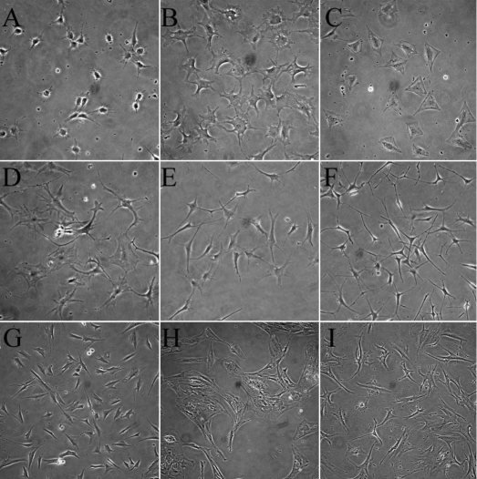
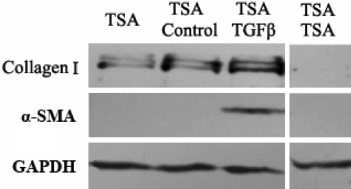
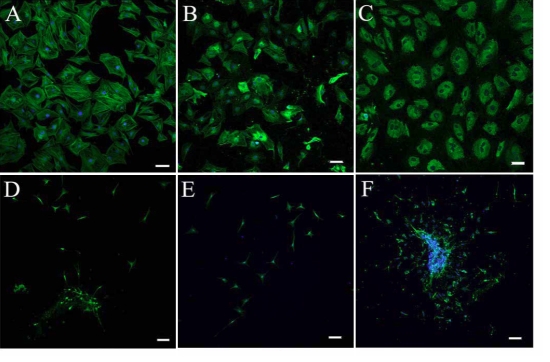
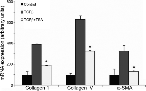
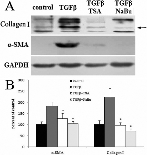
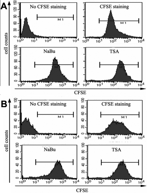
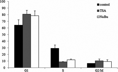
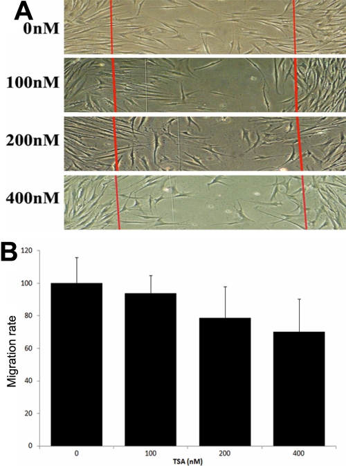
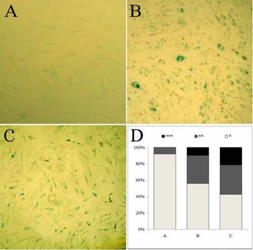
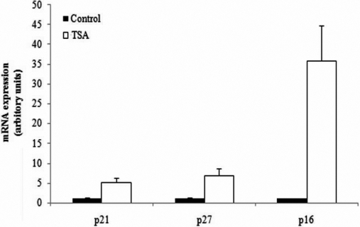
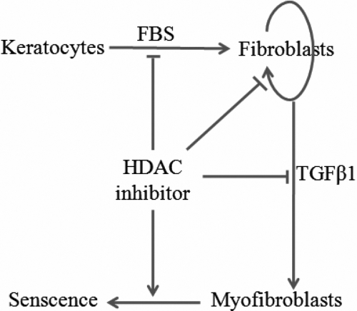
Fetal bovine serum and TGFbeta1 activated the transdifferentiation of corneal stromal cells into fibroblasts and myofibroblasts, indicated by cell spreading, renewed assembly of actin filaments and enhanced expression of extracellular matrix components, all of which were suppressed by the addition of HDAC inhibitors. HDAC inhibitors inhibited the proliferation of corneal fibroblasts by decreasing the proportion in the S-phase and increasing the proportion in the G0/G1 and G2/M cell cycle checkpoints. HDAC inhibitors showed a dose-dependent inhibitory effects on the migration of corneal fibroblasts. In addition, HDAC inhibitors induced the senescence of corneal myofibroblasts as shown by enhanced staining of beta-galactosidase and upregulated expression of p16(ink4a).
HDAC inhibitors may affect corneal stromal cells by inhibiting myofibroblastic differentiation, cell proliferation, migration and by inducing cell senescence. Thus, this has implications for future studies in the development of promising drugs in the prevention or treatment of corneal haze and scar formation.
从活化的角膜基质细胞分化而来的角膜肌成纤维细胞是角膜损伤修复过程中细胞外基质合成的主要细胞来源。在本研究中,评估了组蛋白脱乙酰酶(HDAC)抑制剂对角膜基质细胞的活化、增殖、迁移和衰老的影响。
通过依次用分散酶和胶原酶消化收获原代人及小鼠角膜基质细胞,并在无血清(角膜细胞)、含血清(角膜成纤维细胞)和添加转化生长因子β1(角膜肌成纤维细胞)的条件下于DMEM/F-12培养基中培养。通过cDNA微阵列、实时PCR、免疫细胞化学和蛋白质印迹分析来表征角膜基质细胞对HDAC抑制剂的反应。还在体外评估了HDAC抑制剂对角膜成纤维细胞增殖、细胞周期分布、迁移和衰老的影响。
胎牛血清和转化生长因子β1激活角膜基质细胞向成纤维细胞和肌成纤维细胞的转分化,表现为细胞铺展、肌动蛋白丝重新组装以及细胞外基质成分表达增强,而添加HDAC抑制剂可抑制所有这些现象。HDAC抑制剂通过降低S期比例并增加G0/G1和G2/M细胞周期检查点的比例来抑制角膜成纤维细胞的增殖。HDAC抑制剂对角膜成纤维细胞的迁移表现出剂量依赖性抑制作用。此外,HDAC抑制剂诱导角膜肌成纤维细胞衰老,表现为β-半乳糖苷酶染色增强和p16(ink4a)表达上调。
HDAC抑制剂可能通过抑制肌成纤维细胞分化、细胞增殖、迁移以及诱导细胞衰老来影响角膜基质细胞。因此,这对未来开发用于预防或治疗角膜混浊和瘢痕形成的有前景药物的研究具有启示意义。