Goette Andreas, Bukowska Alicja, Dobrev Dobromir, Pfeiffenberger Jan, Morawietz Henning, Strugala Denis, Wiswedel Ingrid, Röhl Friedrich-Wilhelm, Wolke Carmen, Bergmann Sybille, Bramlage Peter, Ravens Ursula, Lendeckel Uwe
Division of Cardiology, University Hospital Magdeburg, Otto-von-Guericke University, Leipzigerstr. 44, 39120 Magdeburg, Germany.
Eur Heart J. 2009 Jun;30(11):1411-20. doi: 10.1093/eurheartj/ehp046. Epub 2009 Mar 5.
Patients with paroxysmal atrial fibrillation (AF) often present with typical angina pectoris and mildly elevated levels of cardiac troponin (non ST-segment elevation myocardial infarction) during an arrhythmic event. However, in a large proportion of these patients, significant coronary artery disease is excluded by coronary angiography. Here we explored the potential underlying mechanism of these events.
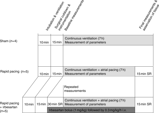
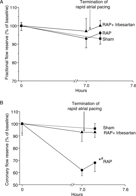
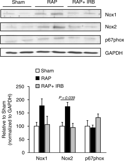
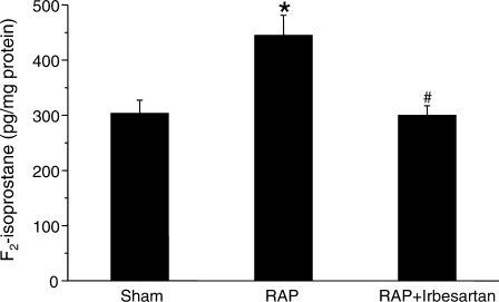
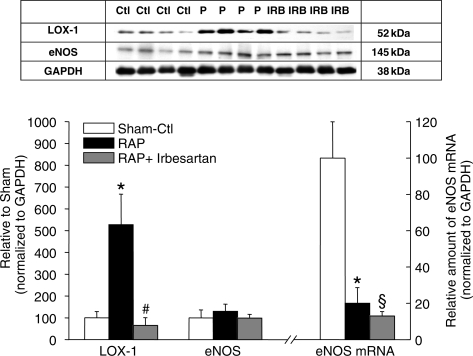
A total of 14 pigs were studied using a closed chest, rapid atrial pacing (RAP) model. In five pigs RAP was performed for 7 h (600 b.p.m.; n = 5), in five animals RAP was performed in the presence of angiotensin-II type-1-receptor (AT(1)-receptor) inhibitor irbesartan (RAP+Irb), and four pigs were instrumented without intervention (Sham). One-factor analysis of variance was performed to assess differences between and within the three groups. Simultaneous measurements of fractional flow reserve (FFR) and coronary flow reserve (CFR) before, during, and after RAP demonstrated unchanged FFR (P = 0.327), but decreased CFR during RAP (RAP: 67.7 +/- 7.2%, sham: 97.2 +/- 2.8%, RAP+Irb: 93.2 +/- 3.3; P = 0.0013) indicating abnormal left ventricular (LV) microcirculation. Alterations in microcirculatory blood flow were accompanied by elevated ventricular expression of NADPH oxidase subunit Nox2 (P = 0.039), lectin-like oxidized low-density lipoprotein receptor-1 (LOX-1, P = 0.004), and F(2)-isoprostane levels (P = 0.008) suggesting RAP-related oxidative stress. Plasma concentrations of cardiac troponin-I (cTn-I) increased in RAP (RAP: 613.3 +/- 125.8 pmol/L vs. sham: 82.5 +/- 12.5 pmol/L; P = 0.013), whereas protein levels of eNOS and LV function remained unchanged. RAP+Irb prevented the increase of Nox2, LOX-1, and F(2)-isoprostanes, and abolished the impairment of microvascular blood flow.
Rapid atrial pacing induces AT(1)-receptor-mediated oxidative stress in LV myocardium that is accompanied by impaired microvascular blood flow and cTn-I release. These findings provide a plausible mechanism for the frequently observed cTn-I elevation accompanied with typical angina pectoris symptoms in patients with paroxysmal AF and normal (non-stenotic) coronary arteries.
阵发性心房颤动(AF)患者在心律失常发作期间常出现典型心绞痛和心肌肌钙蛋白水平轻度升高(非ST段抬高型心肌梗死)。然而,在这些患者中,很大一部分经冠状动脉造影排除了严重冠状动脉疾病。在此,我们探讨了这些事件潜在的潜在机制。
使用封闭胸腔快速心房起搏(RAP)模型对14头猪进行研究。5头猪进行7小时的RAP(600次/分钟;n = 5),5头动物在存在血管紧张素II 1型受体(AT(1)-受体)抑制剂厄贝沙坦的情况下进行RAP(RAP + Irb),4头猪未进行干预作为假手术组(Sham)。进行单因素方差分析以评估三组之间和组内的差异。在RAP之前、期间和之后同时测量血流储备分数(FFR)和冠状动脉血流储备(CFR),结果显示FFR无变化(P = 0.327),但RAP期间CFR降低(RAP:67.7 +/- 7.2%,假手术组:97.2 +/- 2.8%,RAP + Irb:93.2 +/- 3.3;P = 0.0013),表明左心室(LV)微循环异常。微循环血流的改变伴随着心室中NADPH氧化酶亚基Nox2表达升高(P = 0.039)、凝集素样氧化低密度脂蛋白受体-1(LOX-1,P = 0.004)和F(2)-异前列腺素水平升高(P = 0.008),提示与RAP相关的氧化应激。RAP组中心肌肌钙蛋白I(cTn-I)的血浆浓度升高(RAP:613.3 +/- 125.8 pmol/L vs. 假手术组:82.5 +/- 12.5 pmol/L;P = 0.013),而内皮型一氧化氮合酶(eNOS)的蛋白水平和左心室功能保持不变。RAP + Irb可防止Nox2、LOX-1和F(2)-异前列腺素的增加,并消除微血管血流的损害。
快速心房起搏在左心室心肌中诱导AT(1)-受体介导的氧化应激,同时伴有微血管血流受损和cTn-I释放。这些发现为阵发性AF且冠状动脉正常(无狭窄)的患者中经常观察到的cTn-I升高伴典型心绞痛症状提供了一个合理的机制。