Department of Neurosurgery, University of Iowa, Iowa City, Iowa, USA.
J Neurosurg. 2010 Jun;112(6):1301-7. doi: 10.3171/2009.7.JNS09404.
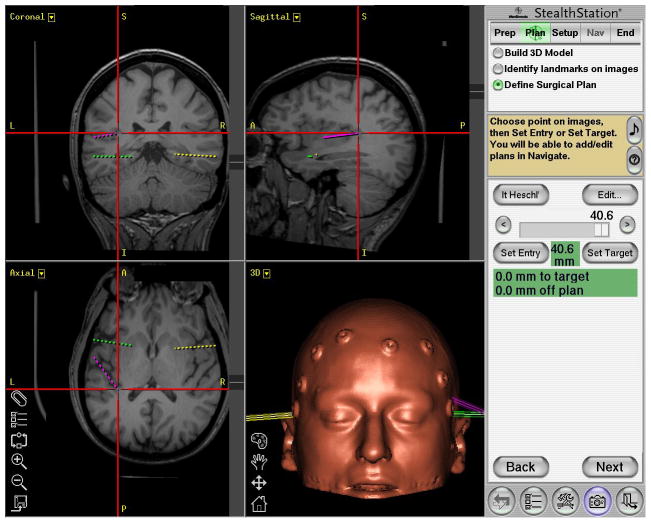
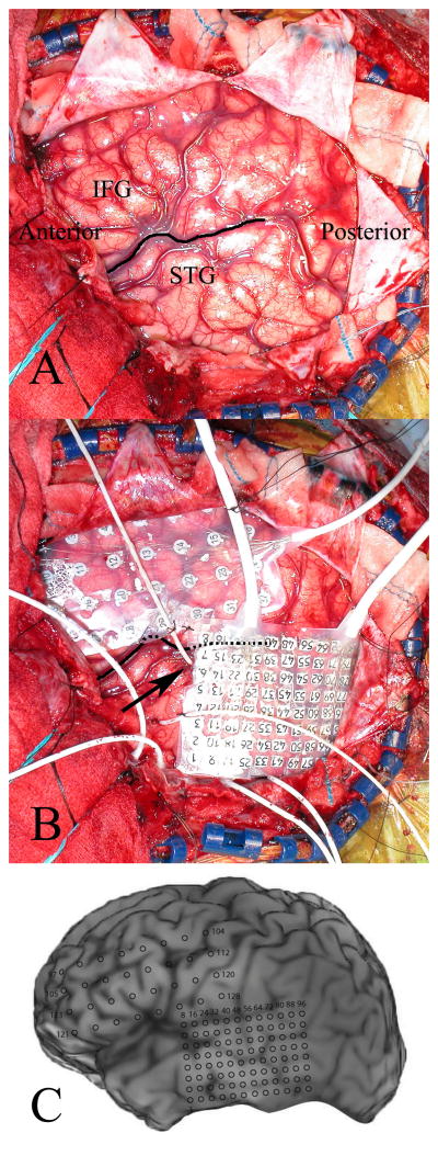
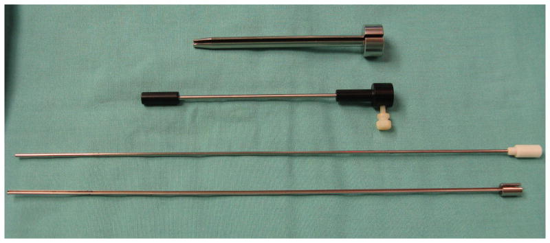
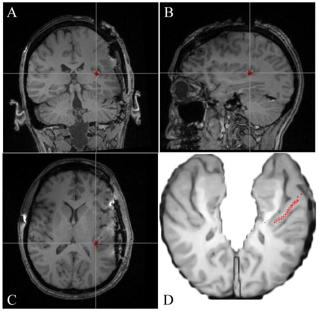
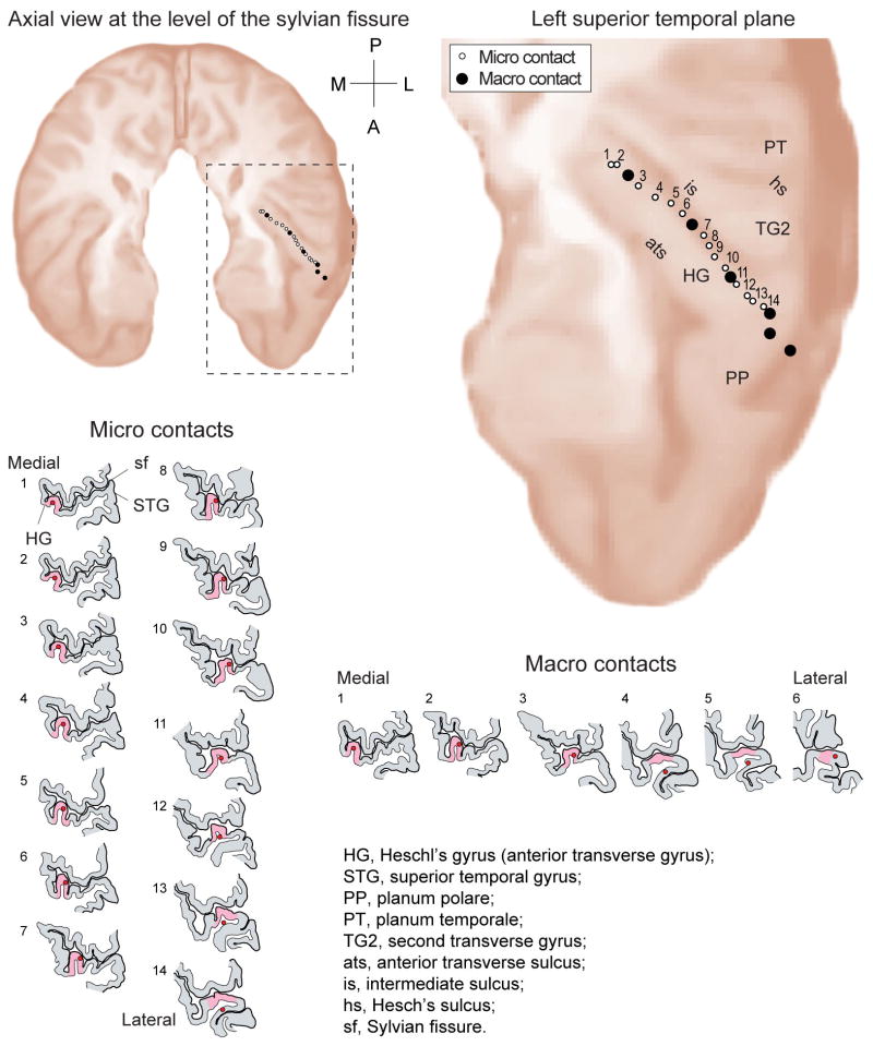
A wide range of devices is used to obtain intracranial electrocorticography recordings in patients with medically refractory epilepsy, including subdural strip and grid electrodes and depth electrodes. Penetrating depth electrodes are required to access some brain regions, and 1 target site that presents a particular technical challenge is the first transverse temporal gyrus, or Heschl gyrus (HG). The HG is located within the supratemporal plane and has an oblique orientation relative to the sagittal and coronal planes. Large and small branches of the middle cerebral artery abut the pial surface of the HG and must be avoided when planning the electrode trajectory. Auditory cortex is located within the HG, and there are functional connections between this dorsal temporal lobe region and medial sites commonly implicated in the pathophysiology of temporal lobe epilepsy. At some surgical centers, depth electrodes are routinely placed within the supratemporal plane, and the HG, in patients who require intracranial electrocorticography monitoring for presumed temporal lobe epilepsy. Information from these recordings is reported to facilitate the identification of seizure patterns in patients with or without auditory auras. To date, only one implantation method has been reported to be safe and effective for placing HG electrodes in a large series of patients undergoing epilepsy surgery. This well-established approach involves inserting the electrodes from a lateral trajectory while using stereoscopic stereotactic angiography to avoid vascular injury. In this report, the authors describe an alternative method for implantation. They use frameless stereotaxy and an oblique insertion trajectory that does not require angiography and allows for the simultaneous placement of subdural grid arrays. Results in 19 patients demonstrate the safety and efficacy of the method.
广泛使用各种设备来获取患有药物难治性癫痫患者的颅内皮质电图记录,包括硬膜下条带和网格电极以及深部电极。需要穿透性深部电极才能进入一些脑区,而一个具有特殊技术挑战的目标区域是第一个横向颞回,或 Heschl 回(HG)。HG 位于颞上平面内,相对于矢状面和冠状面呈倾斜方向。大脑中动脉的大分支和小分支紧贴 HG 的软脑膜表面,在规划电极轨迹时必须避开。听觉皮层位于 HG 内,并且这个背侧颞叶区域与内侧区域之间存在功能连接,内侧区域通常与颞叶癫痫的病理生理学有关。在一些手术中心,在需要颅内皮质电图监测以明确颞叶癫痫的患者中,常规在颞上平面内放置深部电极,以及 HG。这些记录的信息有助于识别有或无听觉先兆的患者的癫痫发作模式。迄今为止,只有一种植入方法在大量接受癫痫手术的患者中被报道是安全有效的,这种方法涉及从外侧轨迹插入电极,同时使用立体定向血管造影术避免血管损伤。在本报告中,作者描述了一种替代的植入方法。他们使用无框架立体定向技术和倾斜插入轨迹,不需要血管造影术,并允许同时放置硬膜下网格阵列。19 例患者的结果证明了该方法的安全性和有效性。