• 文献检索
  • 文档翻译
  • 深度研究
  • 学术资讯
  • Suppr Zotero 插件Zotero 插件
  • 邀请有礼
  • 套餐&价格
  • 历史记录
应用&插件
Suppr Zotero 插件Zotero 插件浏览器插件Mac 客户端Windows 客户端微信小程序
定价
高级版会员购买积分包购买API积分包
服务
文献检索文档翻译深度研究API 文档MCP 服务
关于我们
关于 Suppr公司介绍联系我们用户协议隐私条款
关注我们

Suppr 超能文献

核心技术专利:CN118964589B侵权必究
粤ICP备2023148730 号-1Suppr @ 2026

文献检索

告别复杂PubMed语法,用中文像聊天一样搜索,搜遍4000万医学文献。AI智能推荐,让科研检索更轻松。

立即免费搜索

文件翻译

保留排版,准确专业,支持PDF/Word/PPT等文件格式,支持 12+语言互译。

免费翻译文档

深度研究

AI帮你快速写综述,25分钟生成高质量综述,智能提取关键信息,辅助科研写作。

立即免费体验

Bcl-2 表达随卵巢肿瘤进展而改变:免疫组织化学评估。

Bcl-2 expression is altered with ovarian tumor progression: an immunohistochemical evaluation.

机构信息

Department of Pathology and Cell Biology, University of South Florida, Tampa, FL 33612, USA.

出版信息

J Ovarian Res. 2009 Oct 25;2:16. doi: 10.1186/1757-2215-2-16.

DOI:10.1186/1757-2215-2-16
PMID:19852858
原文链接:https://pmc.ncbi.nlm.nih.gov/articles/PMC2774291/
Abstract

BACKGROUND

Ovarian cancer is the most lethal gynecologic malignancy. The ovarian tumor microenvironment is comprised of tumor cells, surrounding stroma, and circulating lymphocytes, an important component of the immune response, in tumors. Previous reports have shown that the anti-apoptotic protein Bcl-2 is overexpressed in many solid neoplasms, including ovarian cancers, and contributes to neoplastic transformation and drug-resistant disease, resulting in poor clinical outcome. Likewise, studies indicate improved clinical outcome with increased presence of lymphocytes. Therefore, we sought to examine Bcl-2 expression in normal, benign, and cancerous ovarian tissues to determine the potential relationship between epithelial and stromal Bcl-2 expression in conjunction with the presence of lymphocytes for epithelial ovarian tumor progression.

METHODS

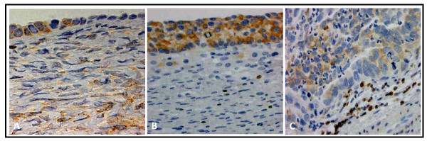
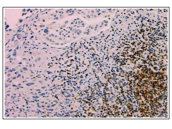
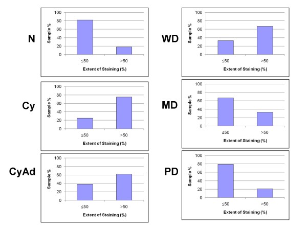
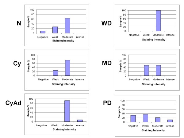
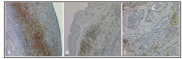
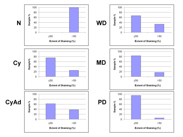
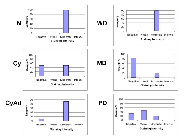
Ovarian tissue sections were classified as normal (n = 2), benign (n = 17) or cancerous (n = 28) and immunohistochemically stained for Bcl-2. Bcl-2 expression was assessed according to cellular localization, extent, and intensity of staining. The number of lymphocyte nests as well as the number of lymphocytes within these nests was counted.

RESULTS

While Bcl-2 staining remained cytoplasmic, both percent and intensity of epithelial and stromal Bcl-2 staining decreased with tumor progression. Further, the number of lymphocyte nests dramatically increased with tumor progression.

CONCLUSION

The data suggest alterations in Bcl-2 expression and lymphocyte infiltration correlate with epithelial ovarian cancer progression. Consequently, Bcl-2 expression and lymphocyte status may be important for prognostic outcome or useful targets for therapeutic intervention.

摘要

背景

卵巢癌是最致命的妇科恶性肿瘤。卵巢肿瘤微环境由肿瘤细胞、周围基质和循环淋巴细胞组成,是肿瘤中免疫反应的重要组成部分。先前的报告表明,抗凋亡蛋白 Bcl-2 在许多实体瘤中过度表达,包括卵巢癌,并有助于肿瘤转化和耐药性疾病,导致临床预后不良。同样,研究表明淋巴细胞的存在增加会改善临床结果。因此,我们试图检查正常、良性和癌性卵巢组织中的 Bcl-2 表达,以确定上皮细胞和基质 Bcl-2 表达与上皮性卵巢肿瘤进展中淋巴细胞的存在之间的潜在关系。

方法

将卵巢组织切片分类为正常(n=2)、良性(n=17)或癌症(n=28),并用 Bcl-2 免疫组织化学染色。根据细胞定位、染色程度和强度评估 Bcl-2 表达。计算淋巴细胞巢的数量以及这些巢内的淋巴细胞数量。

结果

虽然 Bcl-2 染色仍然是细胞质的,但上皮和基质 Bcl-2 染色的百分比和强度随着肿瘤的进展而降低。此外,随着肿瘤的进展,淋巴细胞巢的数量急剧增加。

结论

数据表明 Bcl-2 表达和淋巴细胞浸润的改变与上皮性卵巢癌的进展相关。因此,Bcl-2 表达和淋巴细胞状态可能对预后结果很重要,或者是治疗干预的有用靶点。

https://cdn.ncbi.nlm.nih.gov/pmc/blobs/8765/2774291/38e558c2bed3/1757-2215-2-16-7.jpg
https://cdn.ncbi.nlm.nih.gov/pmc/blobs/8765/2774291/53317a7f6050/1757-2215-2-16-1.jpg
https://cdn.ncbi.nlm.nih.gov/pmc/blobs/8765/2774291/81b7c95f7837/1757-2215-2-16-2.jpg
https://cdn.ncbi.nlm.nih.gov/pmc/blobs/8765/2774291/4a66257e9681/1757-2215-2-16-3.jpg
https://cdn.ncbi.nlm.nih.gov/pmc/blobs/8765/2774291/9bedd7073657/1757-2215-2-16-4.jpg
https://cdn.ncbi.nlm.nih.gov/pmc/blobs/8765/2774291/9c248e85d473/1757-2215-2-16-5.jpg
https://cdn.ncbi.nlm.nih.gov/pmc/blobs/8765/2774291/f37c856cfd39/1757-2215-2-16-6.jpg
https://cdn.ncbi.nlm.nih.gov/pmc/blobs/8765/2774291/38e558c2bed3/1757-2215-2-16-7.jpg

相似文献

1
Bcl-2 expression is altered with ovarian tumor progression: an immunohistochemical evaluation.Bcl-2 表达随卵巢肿瘤进展而改变:免疫组织化学评估。
J Ovarian Res. 2009 Oct 25;2:16. doi: 10.1186/1757-2215-2-16.
2
The clinicopathological significance of Bax and Bcl-2 protein expression with tumor infiltrating lymphocytes in ovarian carcinoma.卵巢癌中 Bax 和 Bcl-2 蛋白表达与肿瘤浸润淋巴细胞的临床病理意义。
Neoplasma. 2012;59(5):475-85. doi: 10.4149/neo_2012_061.
3
Overexpression of epithelial macrophage colony-stimulating factor (CSF-1) and CSF-1 receptor: a poor prognostic factor in epithelial ovarian cancer, contrasted with a protective effect of stromal CSF-1.上皮巨噬细胞集落刺激因子(CSF-1)及其受体的过表达:上皮性卵巢癌的不良预后因素,与基质CSF-1的保护作用形成对比。
Clin Cancer Res. 1997 Jun;3(6):999-1007.
4
Apoptosis and apoptosis-related proteins (Fas, Fas ligand, Blc-2, p53) in lymphoid elements of human ovarian tumors.人卵巢肿瘤淋巴样成分中的细胞凋亡及凋亡相关蛋白(Fas、Fas配体、Bcl-2、p53)
Eur J Gynaecol Oncol. 2000;21(1):53-7.
5
Immunohistochemical analysis of bcl-2, bax, mcl-1, and bcl-X expression in ovarian surface epithelial tumors.卵巢表面上皮性肿瘤中bcl-2、bax、mcl-1和bcl-X表达的免疫组织化学分析
Int J Gynecol Pathol. 1998 Jul;17(3):255-60. doi: 10.1097/00004347-199807000-00010.
6
Significance of apoptosis related proteins on malignant transformation of ovarian tumors: A comparison between Bcl-2/Bax ratio and p53 immunoreactivity.凋亡相关蛋白在卵巢肿瘤恶变中的意义:Bcl-2/Bax比值与p53免疫反应性的比较
Acta Histochem. 2014 Oct;116(8):1251-8. doi: 10.1016/j.acthis.2014.07.007. Epub 2014 Aug 6.
7
Stromal-epithelial interactions in the progression of ovarian cancer: influence and source of tumor stromal cells.卵巢癌进展中的基质-上皮相互作用:肿瘤基质细胞的影响及来源
Mol Cell Endocrinol. 2001 Apr 25;175(1-2):29-39. doi: 10.1016/s0303-7207(01)00436-1.
8
High expression of B7-H3 on stromal cells defines tumor and stromal compartments in epithelial ovarian cancer and is associated with limited immune activation.B7-H3 在基质细胞中的高表达定义了上皮性卵巢癌中的肿瘤和基质区室,并与有限的免疫激活相关。
J Immunother Cancer. 2019 Dec 31;7(1):357. doi: 10.1186/s40425-019-0816-5.
9
Prognostic value of matrix metalloproteinase-9 (gelatinase-B) expression in epithelial ovarian tumors.基质金属蛋白酶-9(明胶酶-B)在上皮性卵巢肿瘤中的表达的预后价值。
Eur J Gynaecol Oncol. 2003;24(5):417-20.
10
Bcl-2 and p53 protein expression, apoptosis, and p53 mutation in human epithelial ovarian cancers.人上皮性卵巢癌中Bcl-2和p53蛋白表达、细胞凋亡及p53突变
Am J Pathol. 2000 Feb;156(2):409-17. doi: 10.1016/S0002-9440(10)64744-X.

引用本文的文献

1
Clinicopathological insights and prognostic implications of DEK in association with apoptosis-regulating factors in ovarian cancer.DEK与卵巢癌中凋亡调节因子相关的临床病理见解及预后意义
Clin Transl Oncol. 2025 May 20. doi: 10.1007/s12094-025-03944-8.
2
Calcineurin Gamma Catalytic Subunit PPP3CC Inhibition by miR-200c-3p Affects Apoptosis in Epithelial Ovarian Cancer.miR-200c-3p 通过抑制钙调神经磷酸酶 γ 催化亚基 PPP3CC 影响上皮性卵巢癌细胞凋亡。
Genes (Basel). 2021 Sep 10;12(9):1400. doi: 10.3390/genes12091400.
3
Nanotechnology-Based Cisplatin Intracellular Delivery to Enhance Chemo-Sensitivity of Ovarian Cancer.

本文引用的文献

1
Intraepithelial T cells and tumor proliferation: impact on the benefit from surgical cytoreduction in advanced serous ovarian cancer.上皮内T细胞与肿瘤增殖:对晚期浆液性卵巢癌手术细胞减灭术获益的影响
Cancer. 2009 Jul 1;115(13):2891-902. doi: 10.1002/cncr.24317.
2
The fallopian tube: primary site of most pelvic high-grade serous carcinomas.输卵管:大多数盆腔高级别浆液性癌的主要发生部位。
Int J Gynecol Cancer. 2009 Jan;19(1):58-64. doi: 10.1111/IGC.0b013e318199009c.
3
Intraepithelial T cells and prognosis in ovarian carcinoma: novel associations with stage, tumor type, and BRCA1 loss.
基于纳米技术的顺铂细胞内递送增强卵巢癌细胞化疗敏感性
Int J Nanomedicine. 2020 Jul 7;15:4793-4810. doi: 10.2147/IJN.S247114. eCollection 2020.
4
Differential expression of immune related genes in high-grade ovarian serous carcinoma.高级别卵巢浆液性癌中免疫相关基因的差异表达。
Gynecol Oncol. 2020 Mar;156(3):662-668. doi: 10.1016/j.ygyno.2019.12.019. Epub 2020 Jan 7.
5
Identification and Validation of Core Genes Involved in the Development of Papillary Thyroid Carcinoma via Bioinformatics Analysis.通过生物信息学分析鉴定和验证参与甲状腺乳头状癌发生发展的核心基因
Int J Genomics. 2019 Sep 8;2019:5894926. doi: 10.1155/2019/5894926. eCollection 2019.
6
Advances in understanding mechanisms of long-term sperm storage-the soft-shelled turtle model.了解长期精子存储机制的进展——软壳龟模型。
Histol Histopathol. 2020 Jan;35(1):1-23. doi: 10.14670/HH-18-148. Epub 2019 Jul 10.
7
Mitochondrially targeted p53 or DBD subdomain is superior to wild type p53 in ovarian cancer cells even with strong dominant negative mutant p53.线粒体靶向的 p53 或 DBD 亚结构域比野生型 p53 在卵巢癌细胞中更有效,即使存在强的显性负突变 p53。
J Ovarian Res. 2019 May 15;12(1):45. doi: 10.1186/s13048-019-0516-2.
8
Impact of the secretome of activated pancreatic stellate cells on growth and differentiation of pancreatic tumour cells.激活的胰腺星状细胞的分泌组对胰腺肿瘤细胞的生长和分化的影响。
Sci Rep. 2019 Mar 28;9(1):5303. doi: 10.1038/s41598-019-41740-x.
9
Catching moving targets: cancer stem cell hierarchies, therapy-resistance & considerations for clinical intervention.捕捉移动目标:癌症干细胞层级结构、治疗抗性及临床干预考量
Mol Cancer. 2017 Feb 23;16(1):43. doi: 10.1186/s12943-017-0601-3.
10
Inhibition of STAT3 signaling as critical molecular event in resveratrol-suppressed ovarian cancer cells.抑制STAT3信号传导作为白藜芦醇抑制卵巢癌细胞的关键分子事件。
J Ovarian Res. 2015 Apr 22;8:25. doi: 10.1186/s13048-015-0152-4.
上皮内T细胞与卵巢癌预后:与分期、肿瘤类型及BRCA1缺失的新关联
Mod Pathol. 2009 Mar;22(3):393-402. doi: 10.1038/modpathol.2008.191. Epub 2008 Dec 5.
4
Urinary levels of Bcl-2 are elevated in ovarian cancer patients.卵巢癌患者尿液中Bcl-2水平升高。
Gynecol Oncol. 2009 Jan;112(1):60-7. doi: 10.1016/j.ygyno.2008.09.037. Epub 2008 Nov 12.
5
Clinical significance of tumor-infiltrating lymphocytes in neoplastic progression and lymph node metastasis of human breast cancer.肿瘤浸润淋巴细胞在人乳腺癌肿瘤进展及淋巴结转移中的临床意义
Breast. 2008 Dec;17(6):604-10. doi: 10.1016/j.breast.2008.06.001. Epub 2008 Jul 24.
6
Ovarian carcinogenesis: an alternative hypothesis.卵巢癌发生:一种替代假说。
Adv Exp Med Biol. 2008;622:79-87. doi: 10.1007/978-0-387-68969-2_7.
7
Cancer statistics, 2008.2008年癌症统计数据。
CA Cancer J Clin. 2008 Mar-Apr;58(2):71-96. doi: 10.3322/CA.2007.0010. Epub 2008 Feb 20.
8
Epithelial-mesenchymal interconversions in normal ovarian surface epithelium and ovarian carcinomas: an exception to the norm.正常卵巢表面上皮及卵巢癌中的上皮-间质转化:一种异常情况
J Cell Physiol. 2007 Dec;213(3):581-8. doi: 10.1002/jcp.21240.
9
Epithelial ovarian cancer: disease etiology, treatment, detection, and investigational gene, metabolite, and protein biomarkers.上皮性卵巢癌:疾病病因、治疗、检测以及研究性基因、代谢物和蛋白质生物标志物
J Proteome Res. 2007 Aug;6(8):2936-62. doi: 10.1021/pr070041v. Epub 2007 Jun 21.
10
Immunohistological characterisation of tumour infiltrating lymphocytes in melanocytic skin lesions.黑素细胞性皮肤病变中肿瘤浸润淋巴细胞的免疫组织学特征
J Clin Pathol. 2006 Mar;59(3):316-24. doi: 10.1136/jcp.2005.028860.