Department of Pharmacology and Toxicology, Rutgers University, Ernest Mario School of Pharmacy, Piscataway, NJ 08854, USA.
Toxicol Appl Pharmacol. 2010 May 15;245(1):36-46. doi: 10.1016/j.taap.2010.01.008. Epub 2010 Jan 25.
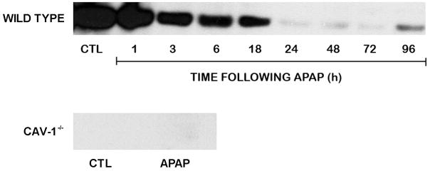
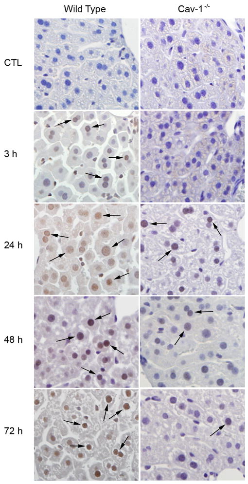
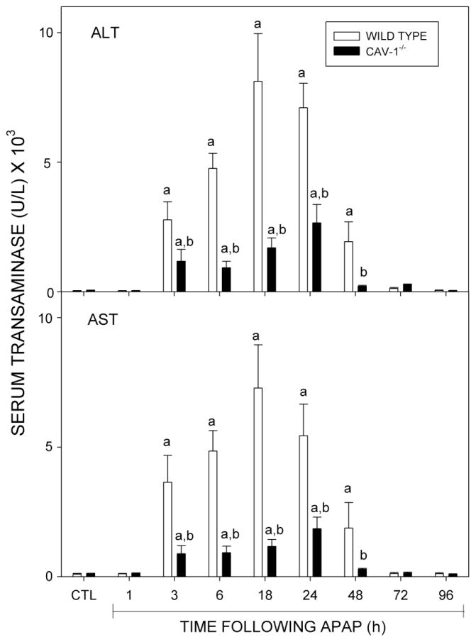
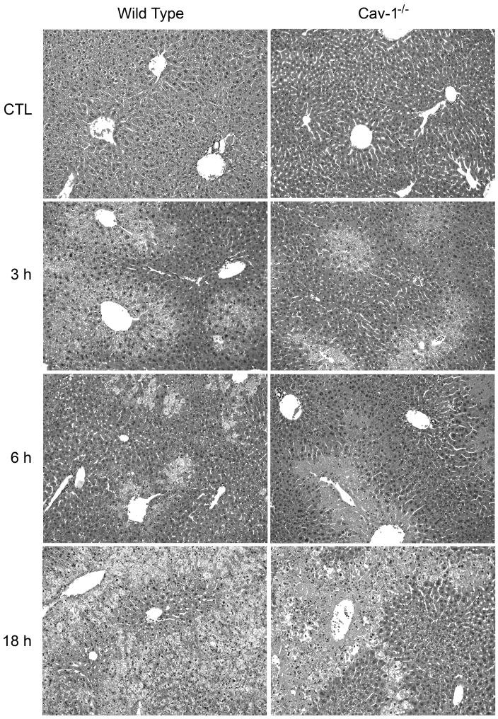
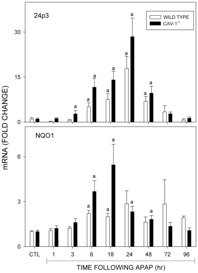
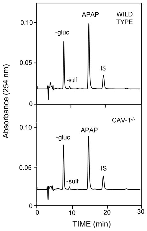
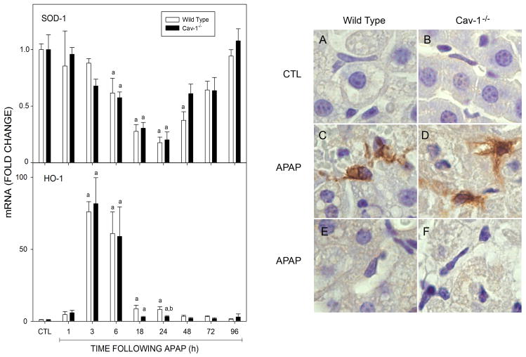
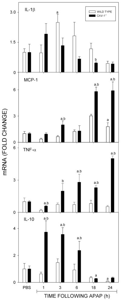
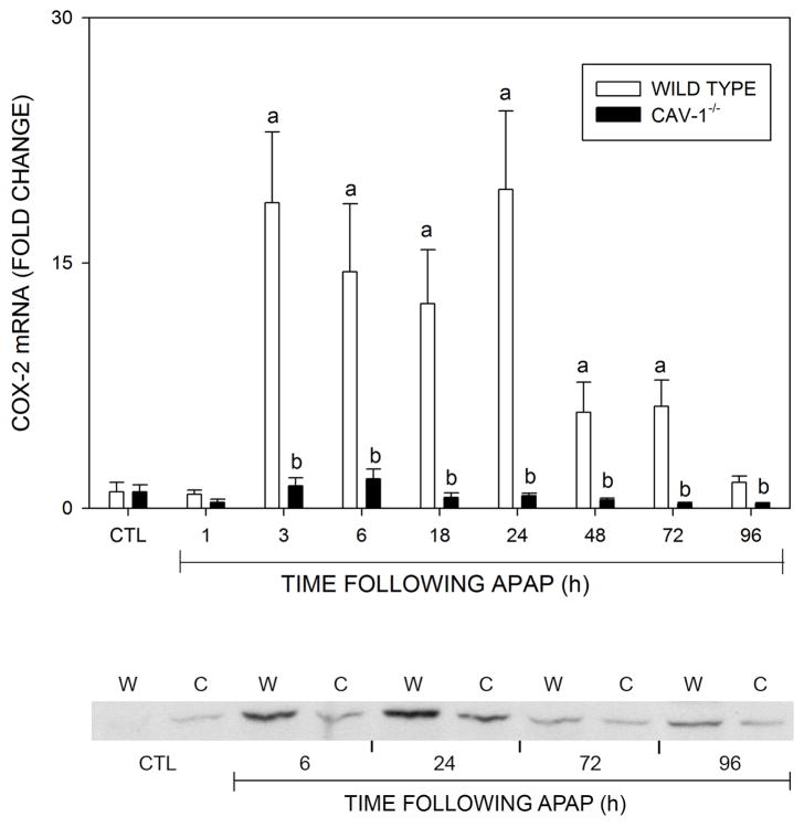
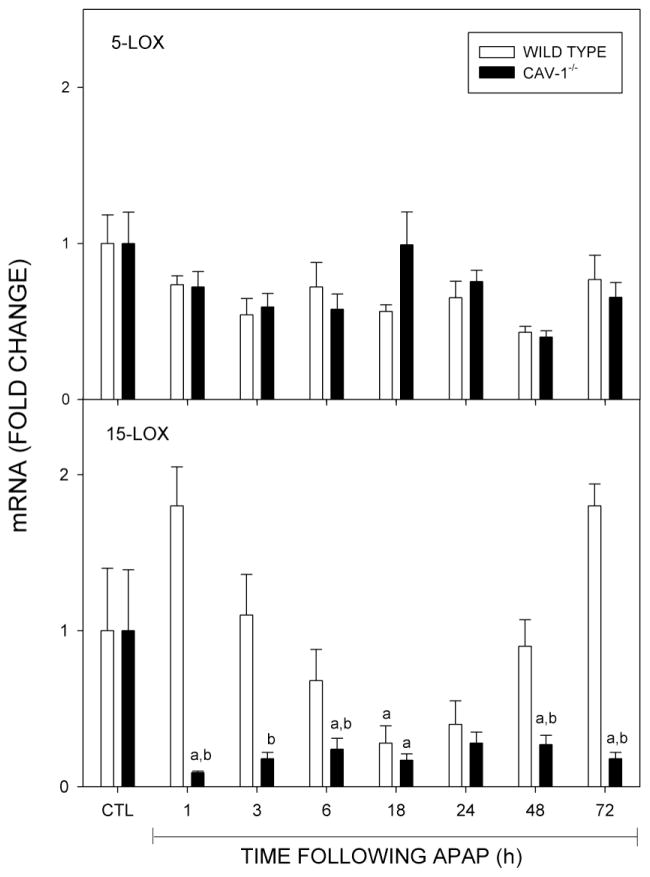
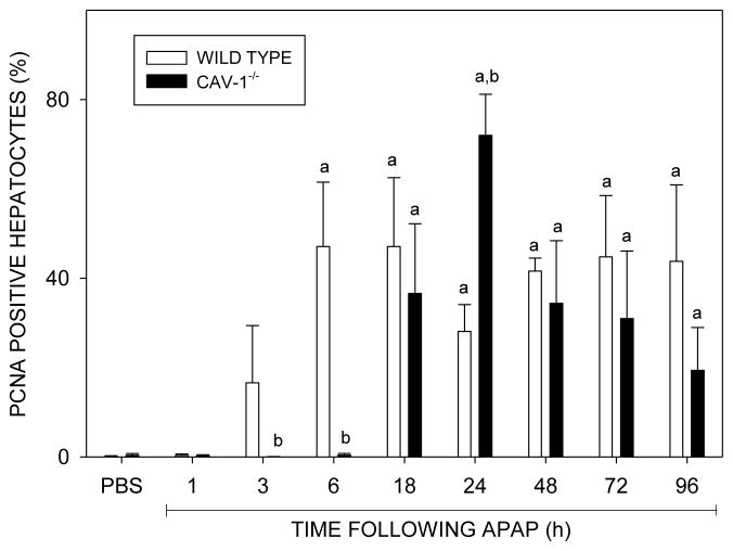
Caveolin-1 (Cav-1) is a membrane scaffolding protein, which functions to regulate intracellular compartmentalization of various signaling molecules. In the present studies, transgenic mice with a targeted disruption of the Cav-1 gene (Cav-1(-/-)) were used to assess the role of Cav-1 in acetaminophen-induced hepatotoxicity. Treatment of wild-type mice with acetaminophen (300 mg/kg) resulted in centrilobular hepatic necrosis and increases in serum transaminases. This was correlated with decreased expression of Cav-1 in the liver. Acetaminophen-induced hepatotoxicity was significantly attenuated in Cav-1(-/-) mice, an effect that was independent of acetaminophen metabolism. Acetaminophen administration resulted in increased hepatic expression of the oxidative stress marker, lipocalin 24p3, as well as hemeoxygenase-1, but decreased glutathione and superoxide dismutase-1; no differences were noted between the genotypes suggesting that reduced toxicity in Cav-1(-/-) mice is not due to alterations in antioxidant defense. In wild-type mice, acetaminophen increased mRNA expression of the pro-inflammatory cytokines, interleukin-1beta, and monocyte chemoattractant protein-1 (MCP-1), as well as cyclooxygenase-2, while 15-lipoxygenase (15-LOX), which generates anti-inflammatory lipoxins, decreased. Acetaminophen-induced changes in MCP-1 and 15-LOX expression were greater in Cav-1(-/-) mice. Although expression of tumor necrosis factor-alpha, a potent hepatocyte mitogen, was up-regulated in the liver of Cav-1(-/-) mice after acetaminophen, expression of proliferating cell nuclear antigen and survivin, markers of cellular proliferation, were delayed, which may reflect the reduced need for tissue repair. Taken together, these data demonstrate that Cav-1 plays a role in promoting inflammation and toxicity during the pathogenesis of acetaminophen-induced injury.
窖蛋白-1(Cav-1)是一种膜支架蛋白,其功能是调节各种信号分子的细胞内区室化。在本研究中,使用靶向敲除 Cav-1 基因(Cav-1(-/-))的转基因小鼠来评估 Cav-1 在对乙酰氨基酚诱导的肝毒性中的作用。用对乙酰氨基酚(300mg/kg)处理野生型小鼠导致中央小叶肝坏死和血清转氨酶升高。这与肝中 Cav-1 的表达减少有关。在 Cav-1(-/-)小鼠中,对乙酰氨基酚诱导的肝毒性明显减弱,这种作用与对乙酰氨基酚代谢无关。对乙酰氨基酚给药导致肝中氧化应激标志物脂联素 24p3 和血红素加氧酶-1 的表达增加,但谷胱甘肽和超氧化物歧化酶-1 的表达减少;基因型之间没有差异,表明 Cav-1(-/-)小鼠的毒性降低不是由于抗氧化防御的改变。在野生型小鼠中,对乙酰氨基酚增加了促炎细胞因子白细胞介素-1β和单核细胞趋化蛋白-1(MCP-1)以及环氧化酶-2的 mRNA 表达,而生成抗炎脂氧素的 15-脂氧合酶(15-LOX)减少。在 Cav-1(-/-)小鼠中,对乙酰氨基酚诱导的 MCP-1 和 15-LOX 表达的变化更大。尽管在 Cav-1(-/-)小鼠的肝中,对乙酰氨基酚后肿瘤坏死因子-α(一种有效的肝细胞有丝分裂原)的表达上调,但细胞增殖的标志物增殖细胞核抗原和存活素的表达延迟,这可能反映出组织修复的需求减少。总之,这些数据表明 Cav-1 在促进对乙酰氨基酚诱导损伤发病机制中的炎症和毒性方面发挥作用。