Division of Cardiovascular Diseases, Department of Internal Medicine, Mayo Clinic, 200 First Street SW, Rochester, MN 55905, USA.
Circ Res. 2010 Apr 2;106(6):1164-73. doi: 10.1161/CIRCRESAHA.109.209767. Epub 2010 Feb 18.
The large conductance Ca(2+)-activated K(+) (BK) channel, a key determinant of vascular tone, is regulated by angiotensin II (Ang II) type 1 receptor signaling. Upregulation of Ang II functions and downregulation of BK channel activities have been reported in diabetic vessels. However, the molecular mechanisms underlying Ang II-mediated BK channel modulation, especially in diabetes mellitus, have not been thoroughly examined.
The aim in this study was to determine whether caveolae-targeting facilitates BK channel dysfunction in diabetic vessels.
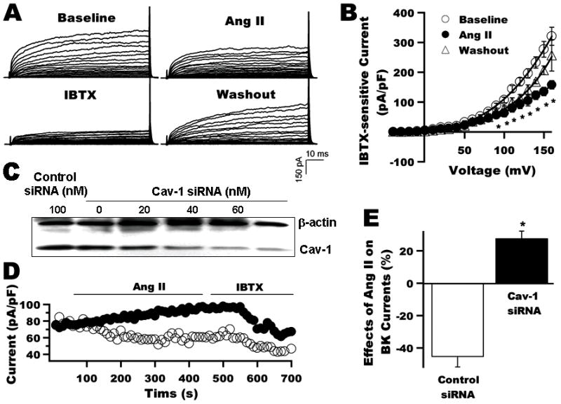
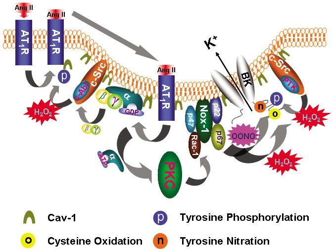
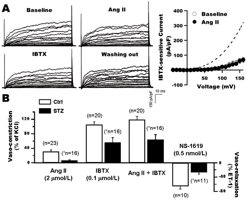
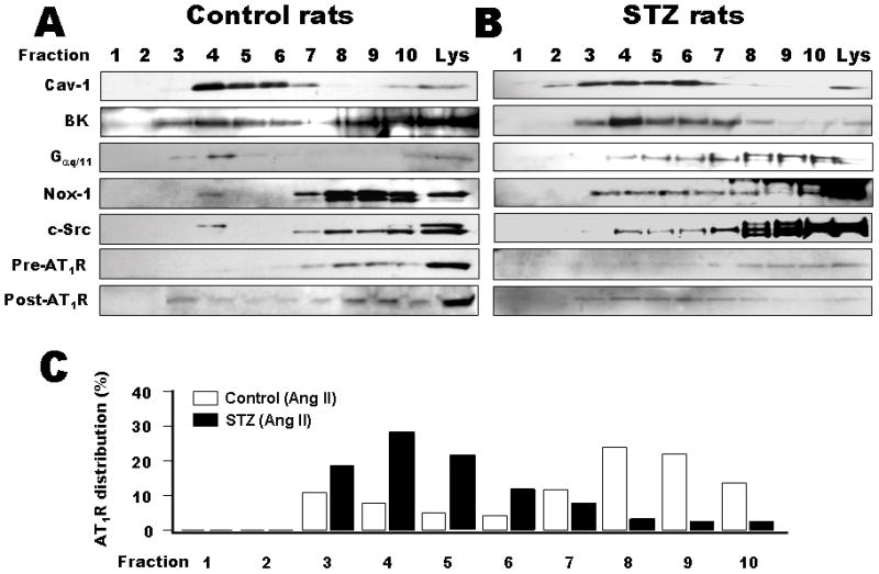
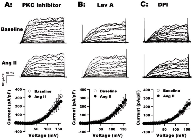
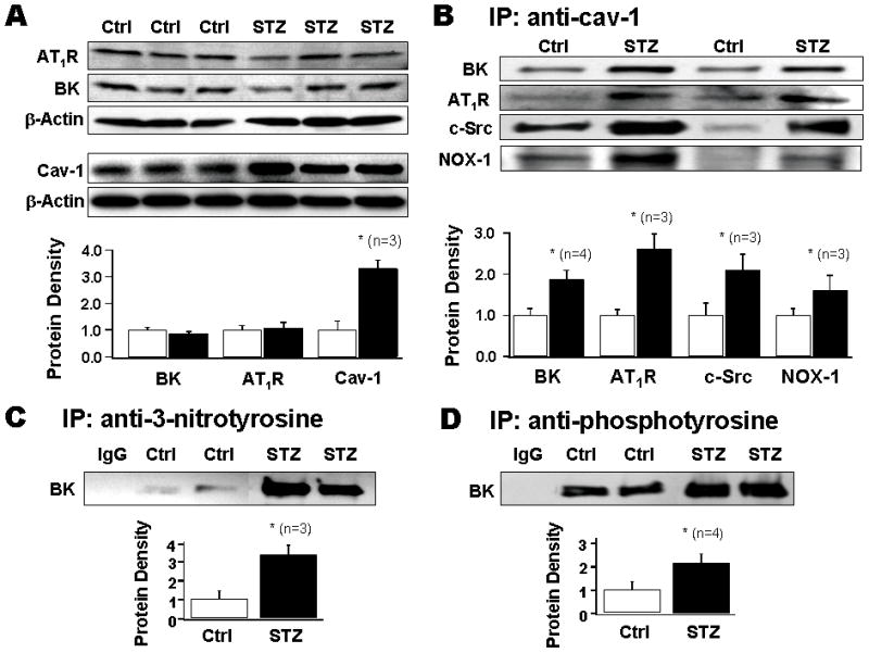
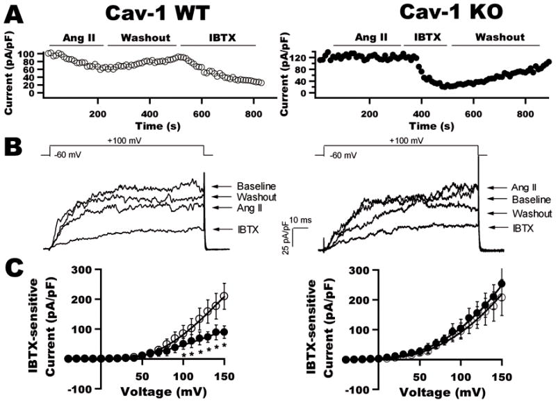
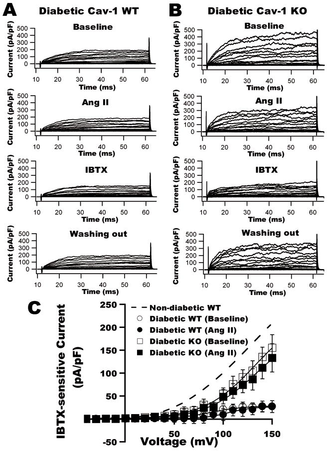
Using patch clamp techniques and molecular biological approaches, we found that BK channels, Ang II type 1 receptor, G(alphaq/11) (G protein q/11 alpha subunit), nonphagocytic NAD(P)H oxidases (NOX-1), and c-Src kinases (c-Src) were colocalized in the caveolae of rat arterial smooth muscle cells and the integrity of caveolae in smooth muscle cells was critical for Ang II-mediated BK channel regulation. Most importantly, membrane microdomain targeting of these proteins was upregulated in the caveolae of streptozotocin-induced rat diabetic vessels, leading to enhanced Ang II-induced redox-mediated BK channel modification and causing BK channel and coronary dysfunction. The absence of caveolae abolished the effects of Ang II on vascular BK channel activity and preserved BK channel function in diabetes.
These results identified a molecular scheme of receptor/enzyme/channel/caveolae microdomain complex that facilitates the development of vascular BK channel dysfunction in diabetes.
大电导钙激活钾(BK)通道是血管张力的关键决定因素,受血管紧张素 II(Ang II)型 1 受体信号转导调节。糖尿病血管中 Ang II 功能上调和 BK 通道活性下调已有报道。然而,Ang II 介导的 BK 通道调节的分子机制,特别是在糖尿病中,尚未得到彻底研究。
本研究旨在确定是否靶向质膜小窝有利于糖尿病血管中的 BK 通道功能障碍。
使用膜片钳技术和分子生物学方法,我们发现 BK 通道、Ang II 型 1 受体、G(alphaq/11)(G 蛋白 q/11 alpha 亚基)、非吞噬性 NAD(P)H 氧化酶(NOX-1)和 c-Src 激酶(c-Src)在大鼠动脉平滑肌细胞的质膜小窝中存在共定位,并且质膜小窝中平滑肌细胞的质膜小窝完整性对于 Ang II 介导的 BK 通道调节至关重要。最重要的是,这些蛋白在链脲佐菌素诱导的大鼠糖尿病血管中质膜小窝的膜微区靶向作用上调,导致增强的 Ang II 诱导的氧化还原介导的 BK 通道修饰,并导致 BK 通道和冠状功能障碍。质膜小窝的缺失消除了 Ang II 对血管 BK 通道活性的影响,并在糖尿病中保留了 BK 通道功能。
这些结果确定了一种受体/酶/通道/质膜小窝微区复合物的分子方案,该方案促进了糖尿病中血管 BK 通道功能障碍的发展。