Montreal Heart Institute, 5000 Belanger St., Montréal, Québec, Canada.
Cell Signal. 2010 Jul;22(7):1063-75. doi: 10.1016/j.cellsig.2010.02.009. Epub 2010 Mar 7.
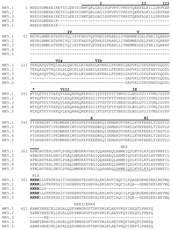
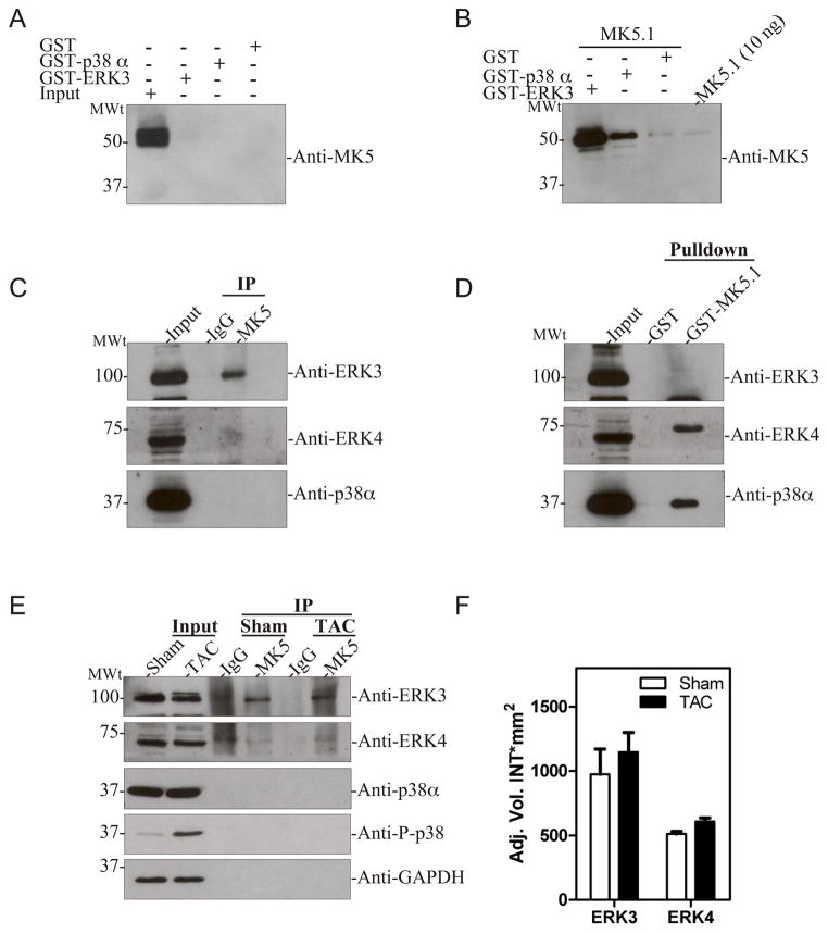
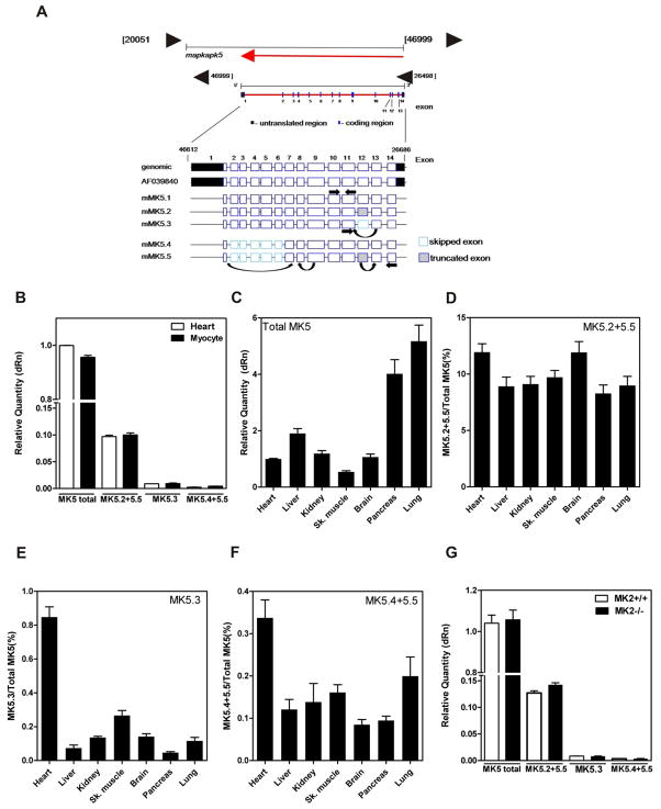
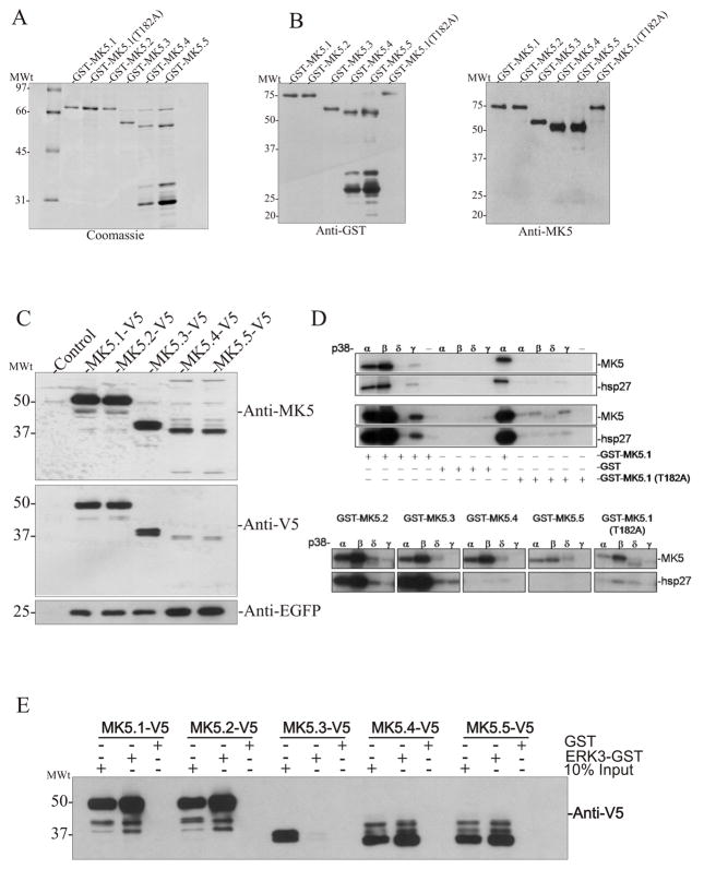
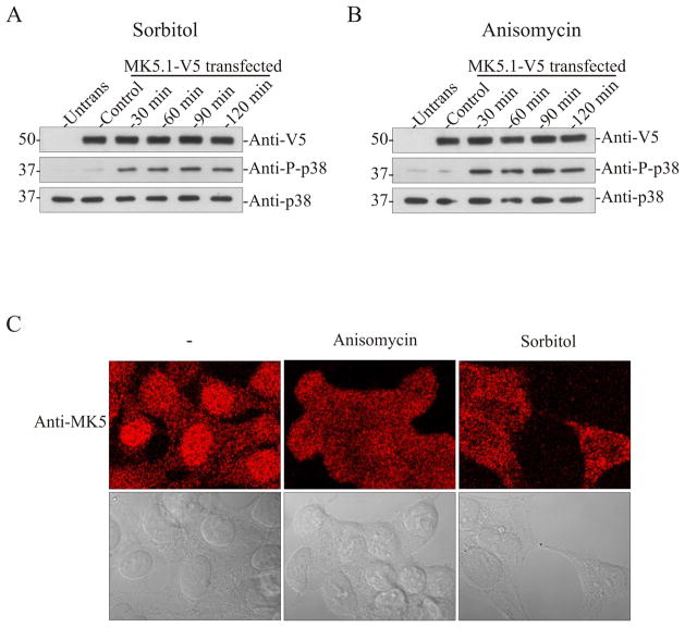
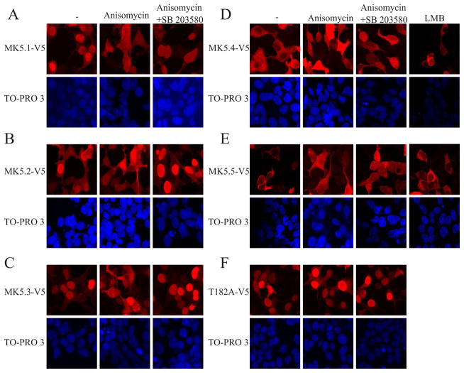
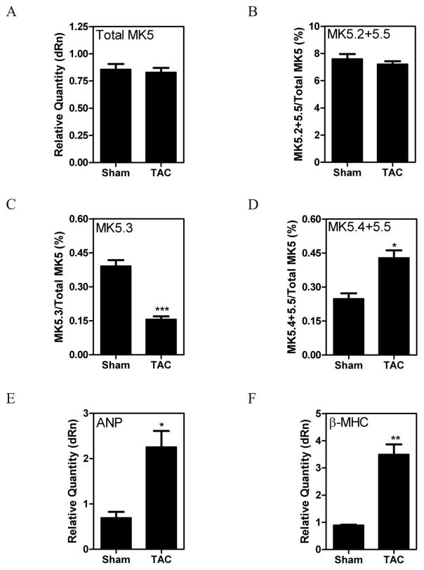
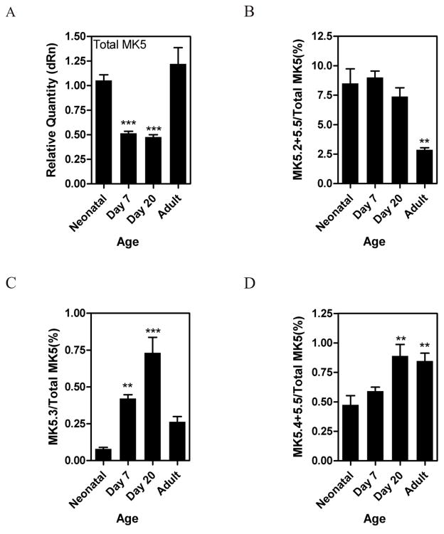
MK5, a member of the MAPK-activated protein kinase family, is highly expressed in the heart. Whereas MK2 and MK3 are activated by p38 MAPK, MK5 has also been shown to be activated by ERK3 and ERK4. We studied the regulation of MK5 in mouse heart. mRNA for 5 splice variants (MK5.1-5.5), including the original form (MK5.1), was detected. MK5 comprises 14 exons: exon 12 splicing was modified in MK5.2, MK5.3, and MK5.5. MK5.2 and MK5.5 lacked 6 bases at the 3'-end of exon 12, whereas MK5.3 lacked exon 12, resulting in a frame shift and premature termination of translation at codon 3 of exon 13. MK5.4 and MK5.5 lacked exons 2-6, encoding kinase subdomains I-VI, and were kinase-dead. All 5 MK5 variants were detected at the mRNA level in all mouse tissues examined; however, their relative abundance was tissue-specific. Furthermore, the relative abundance of variant mRNA was altered both during hypertrophy and postnatal cardiac development, suggesting that the generation or the stability of MK5 variant mRNAs is subject to regulation. When expressed in HEK293 cells, MK5.1, MK5.2 and MK5.3 were nuclear whereas MK5.4 and MK5.5 were cytoplasmic. A p38 MAPK activator, anisomycin, induced the redistribution of each variant. In contrast, MK5 co-immunoprecipitated ERK3, but not ERK4 or p38 alpha, in control and hypertrophying hearts. GST pull-down assays revealed unbound ERK4 and p38 alpha but no free MK5 or ERK3 in heart lysates. Hence, 1) in heart MK5 complexes with ERK3 and 2) MK5 splice variants may mediate distinct effects thus increasing the functional diversity of ERK3-MK5 signaling.
MK5 是丝裂原活化蛋白激酶(MAPK)激活的蛋白激酶家族的成员,在心脏中高度表达。虽然 MK2 和 MK3 被 p38 MAPK 激活,但 MK5 也被 ERK3 和 ERK4 激活。我们研究了小鼠心脏中 MK5 的调节。检测到 5 种剪接变体(MK5.1-5.5)的 mRNA,包括原始形式(MK5.1)。MK5 由 14 个外显子组成:外显子 12 的剪接在 MK5.2、MK5.3 和 MK5.5 中发生了改变。MK5.2 和 MK5.5 在外显子 12 的 3'末端缺少 6 个碱基,而 MK5.3 则缺少外显子 12,导致翻译在第 13 外显子的第 3 位发生移码和提前终止。MK5.4 和 MK5.5 缺失了编码激酶亚结构域 I-VI 的外显子 2-6,是激酶失活的。在检查的所有小鼠组织中均在 mRNA 水平检测到所有 5 种 MK5 变体;然而,它们的相对丰度是组织特异性的。此外,变体 mRNA 的相对丰度在肥大和出生后心脏发育过程中均发生了改变,这表明 MK5 变体 mRNA 的产生或稳定性受到调节。当在 HEK293 细胞中表达时,MK5.1、MK5.2 和 MK5.3 是核内的,而 MK5.4 和 MK5.5 是细胞质的。p38 MAPK 激活剂anisomycin 诱导了每种变体的重新分布。相反,在对照和肥大的心脏中,MK5 与 ERK3 共免疫沉淀,但不与 ERK4 或 p38α共免疫沉淀。GST 下拉测定显示在心脏裂解物中没有游离的 ERK4 和 p38α,但有结合的 ERK3 和 MK5。因此,1)在心脏中,MK5 与 ERK3 结合,2)MK5 剪接变体可能介导不同的作用,从而增加 ERK3-MK5 信号传导的功能多样性。