Department of Orthopaedic Surgery, University of Amsterdam, Academic Medical Center, PO Box 22700, 1100 DE Amsterdam, The Netherlands.
Knee Surg Sports Traumatol Arthrosc. 2010 May;18(5):612-7. doi: 10.1007/s00167-010-1099-z. Epub 2010 Mar 12.
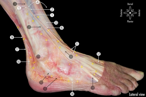
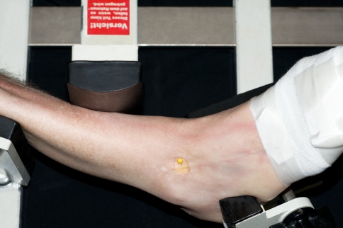
Despite the fact that the superficial peroneal nerve is the only nerve in the human body that can be made visible; iatrogenic damage to this nerve is the most frequently reported complication in anterior ankle arthroscopy. One of the methods to visualize the nerve is combined ankle plantar flexion and inversion. In the majority of cases, the superficial peroneal nerve can be made visible. The portals for anterior ankle arthroscopy are however created with the ankle in the neutral or slightly dorsiflexed position and not in combined plantar flexion and inversion. The purpose of this study was to undertake an anatomical study to the course of the superficial peroneal nerve in different positions of the foot and ankle. We hypothesize that the anatomical localization of the superficial peroneal nerve changes with different foot and ankle positions. In ten fresh frozen ankle specimens, a window, only affecting the skin, was made at the level of the anterolateral portal for anterior ankle arthroscopy in order to directly visualize the superficial peroneal nerve, or if divided, its terminal branches. Nerve movement was assessed from combined 10 degrees plantar flexion and inversion to 5 degrees dorsiflexion, standardized by the Telos stress device. Also for the 4th toe flexion, flexion of all the toes and for skin tensioning possible nerve movement was determined. The mean superficial peroneal nerve movement was 2.4 mm to the lateral side when the ankle was moved from 10 degrees plantar flexion and inversion to the neutral ankle position and 3.6 mm to the lateral side from 10 degrees plantar flexion and inversion to 5 degrees dorsiflexion. Both displacements were significant (P < 0.01). The nerve consistently moves lateral when the ankle is manoeuvred from combined plantar flexion and inversion to the neutral or dorsiflexed position. If visible, it is therefore advised to create the anterolateral portal medial from the preoperative marking, in order to prevent iatrogenic damage to the superficial peroneal nerve.
尽管人体中唯一可见的神经是浅表腓浅神经;但在前踝关节镜检查中,该神经最常报告的并发症是医源性损伤。可见神经的一种方法是联合踝关节跖屈和内翻。在大多数情况下,浅表腓浅神经可以被看见。但是,在前踝关节镜检查的入路是在踝关节中立或轻度背屈的位置而不是在联合跖屈和内翻的位置创建的。本研究的目的是对足部和踝关节不同位置下浅表腓浅神经的走行进行解剖学研究。我们假设解剖学定位的浅表腓浅神经随着足部和踝关节位置的不同而发生变化。在十个新鲜冷冻的踝关节标本中,在用于前踝关节镜检查的前外侧入路水平处制作一个仅影响皮肤的窗口,以便直接观察浅表腓浅神经,或如果已经被切开,则观察其终末分支。使用 Telos 应力装置评估神经运动,从联合 10 度跖屈和内翻到 5 度背屈进行标准化。还评估了 4 趾屈曲、所有脚趾的屈曲和皮肤紧张时可能的神经运动。当踝关节从 10 度跖屈和内翻到中立踝关节位置时,浅表腓浅神经向外侧移动 2.4 毫米,从 10 度跖屈和内翻到 5 度背屈时向外侧移动 3.6 毫米。这两种移位均有统计学意义(P < 0.01)。当踝关节从联合跖屈和内翻到中立或背屈位置时,神经总是向外侧移位。如果可以看见,建议在术前标记的内侧创建前外侧入路,以防止医源性损伤浅表腓浅神经。