• 文献检索
  • 文档翻译
  • 深度研究
  • 学术资讯
  • Suppr Zotero 插件Zotero 插件
  • 邀请有礼
  • 套餐&价格
  • 历史记录
应用&插件
Suppr Zotero 插件Zotero 插件浏览器插件Mac 客户端Windows 客户端微信小程序
定价
高级版会员购买积分包购买API积分包
服务
文献检索文档翻译深度研究API 文档MCP 服务
关于我们
关于 Suppr公司介绍联系我们用户协议隐私条款
关注我们

Suppr 超能文献

核心技术专利:CN118964589B侵权必究
粤ICP备2023148730 号-1Suppr @ 2026

文献检索

告别复杂PubMed语法,用中文像聊天一样搜索,搜遍4000万医学文献。AI智能推荐,让科研检索更轻松。

立即免费搜索

文件翻译

保留排版,准确专业,支持PDF/Word/PPT等文件格式,支持 12+语言互译。

免费翻译文档

深度研究

AI帮你快速写综述,25分钟生成高质量综述,智能提取关键信息,辅助科研写作。

立即免费体验

绵羊关节软骨中胶原蛋白结构的出生后发育

Postnatal development of collagen structure in ovine articular cartilage.

作者信息

van Turnhout Mark C, Schipper Henk, Engel Bas, Buist Willem, Kranenbarg Sander, van Leeuwen Johan L

机构信息

Wageningen University, Department of Animal Sciences, Experimental Zoology Group, 6700 AH Wageningen, the Netherlands.

出版信息

BMC Dev Biol. 2010 Jun 7;10:62. doi: 10.1186/1471-213X-10-62.

DOI:10.1186/1471-213X-10-62
PMID:20529268
原文链接:https://pmc.ncbi.nlm.nih.gov/articles/PMC2906441/
Abstract

BACKGROUND

Articular cartilage (AC) is the layer of tissue that covers the articulating ends of the bones in diarthrodial joints. Across species, adult AC shows an arcade-like structure with collagen predominantly perpendicular to the subchondral bone near the bone, and collagen predominantly parallel to the articular surface near the articular surface. Recent studies into collagen fibre orientation in stillborn and juvenile animals showed that this structure is absent at birth. Since the collagen structure is an important factor for AC mechanics, the absence of the adult Benninghoff structure has implications for perinatal AC mechanobiology. The current objective is to quantify the dynamics of collagen network development in a model animal from birth to maturity. We further aim to show the presence or absence of zonal differentiation at birth, and to assess differences in collagen network development between different anatomical sites of a single joint surface. We use quantitative polarised light microscopy to investigate properties of the collagen network and we use the sheep (Ovis aries) as our model animal.

RESULTS

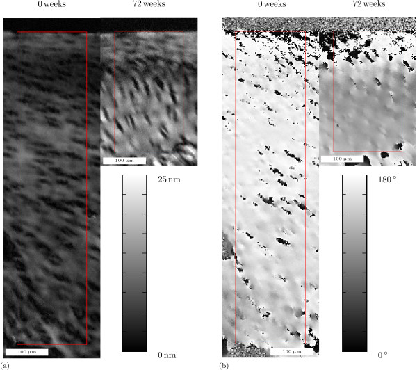
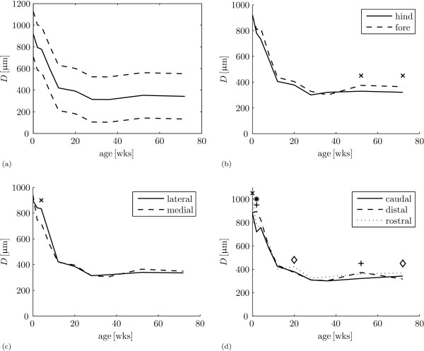
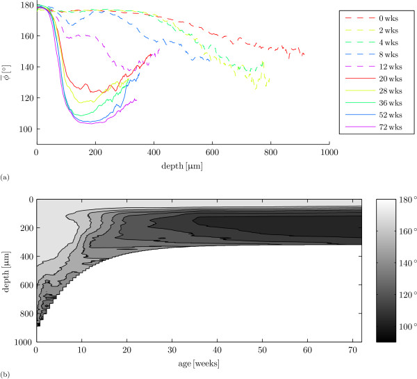
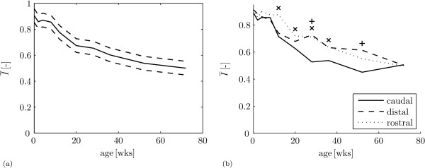
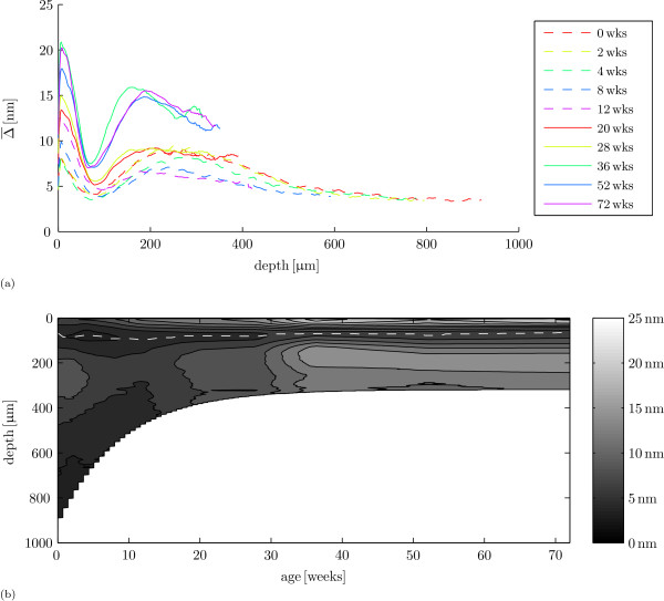
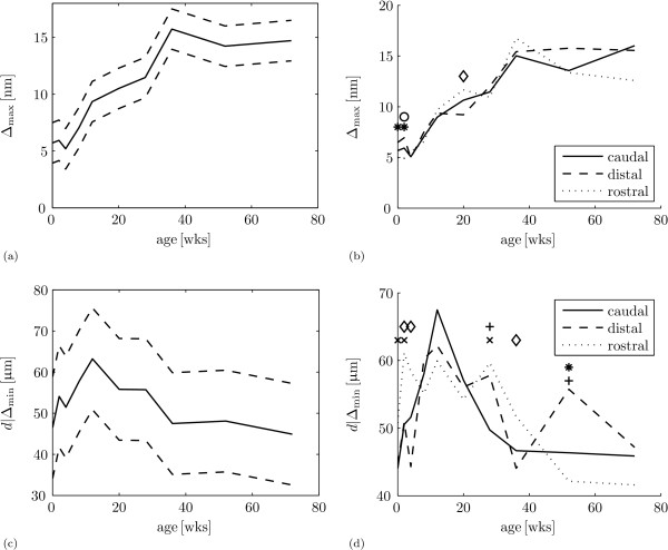
Predominant collagen orientation is parallel to the articular surface throughout the tissue depth for perinatal cartilage. This remodels to the Benninghoff structure before the sheep reach sexual maturity. Remodelling of predominant collagen orientation starts at a depth just below the future transitional zone. Tissue retardance shows a minimum near the articular surface at all ages, which indicates the presence of zonal differentiation at all ages. The absolute position of this minimum does change between birth and maturity. Between different anatomical sites, we find differences in the dynamics of collagen remodelling, but no differences in adult collagen structure.

CONCLUSIONS

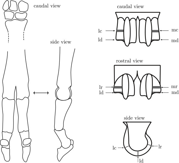
The collagen network in articular cartilage remodels between birth and sexual maturity from a network with predominant orientation parallel to the articular surface to a Benninghoff network. The retardance minimum near, but not at, the articular surface at all ages shows that a zonal differentiation is already present in the perinatal animals. In these animals, the zonal differentiation can not be correlated to the collagen network orientation. We find no difference in adult collagen structure in the nearly congruent metacarpophalangeal joint, but we do find differences in the dynamics of collagen network remodelling.

摘要

背景

关节软骨(AC)是覆盖在滑膜关节骨的关节端的一层组织。在不同物种中,成年AC呈现出一种拱廊状结构,在靠近骨的部位,胶原蛋白主要垂直于软骨下骨,而在靠近关节面的部位,胶原蛋白主要平行于关节面。最近对死产和幼年动物胶原蛋白纤维取向的研究表明,这种结构在出生时并不存在。由于胶原蛋白结构是影响AC力学性能的一个重要因素,成年本宁霍夫结构的缺失对围产期AC的力学生物学具有重要意义。当前的目标是量化模型动物从出生到成熟过程中胶原蛋白网络发育的动态变化。我们进一步旨在揭示出生时是否存在区域分化,并评估单个关节表面不同解剖部位之间胶原蛋白网络发育的差异。我们使用定量偏振光显微镜来研究胶原蛋白网络的特性,并以绵羊(Ovis aries)作为我们的模型动物。

结果

围产期软骨在整个组织深度内,主要胶原蛋白取向平行于关节面。在绵羊达到性成熟之前,这种结构会重塑为本宁霍夫结构。主要胶原蛋白取向的重塑始于未来过渡区下方的深度。组织延迟在所有年龄段都在关节面附近显示出最小值,这表明在所有年龄段都存在区域分化。这个最小值的绝对位置在出生和成熟之间确实会发生变化。在不同解剖部位之间,我们发现胶原蛋白重塑的动态变化存在差异,但成年胶原蛋白结构没有差异。

结论

关节软骨中的胶原蛋白网络在出生到性成熟之间从主要取向平行于关节面的网络重塑为本宁霍夫网络。在所有年龄段,靠近但不在关节面处的延迟最小值表明围产期动物中已经存在区域分化。在这些动物中,区域分化与胶原蛋白网络取向无关。我们发现几乎全等的掌指关节中成年胶原蛋白结构没有差异,但我们确实发现胶原蛋白网络重塑的动态变化存在差异。

https://cdn.ncbi.nlm.nih.gov/pmc/blobs/583f/2906441/85922bc8a9e8/1471-213X-10-62-8.jpg
https://cdn.ncbi.nlm.nih.gov/pmc/blobs/583f/2906441/3aa10387aede/1471-213X-10-62-1.jpg
https://cdn.ncbi.nlm.nih.gov/pmc/blobs/583f/2906441/1a39d15207f4/1471-213X-10-62-2.jpg
https://cdn.ncbi.nlm.nih.gov/pmc/blobs/583f/2906441/801661dcffa4/1471-213X-10-62-3.jpg
https://cdn.ncbi.nlm.nih.gov/pmc/blobs/583f/2906441/8918af582faf/1471-213X-10-62-4.jpg
https://cdn.ncbi.nlm.nih.gov/pmc/blobs/583f/2906441/fb8453533289/1471-213X-10-62-5.jpg
https://cdn.ncbi.nlm.nih.gov/pmc/blobs/583f/2906441/f5eb06d5a412/1471-213X-10-62-6.jpg
https://cdn.ncbi.nlm.nih.gov/pmc/blobs/583f/2906441/4502c85155a0/1471-213X-10-62-7.jpg
https://cdn.ncbi.nlm.nih.gov/pmc/blobs/583f/2906441/85922bc8a9e8/1471-213X-10-62-8.jpg

相似文献

1
Postnatal development of collagen structure in ovine articular cartilage.绵羊关节软骨中胶原蛋白结构的出生后发育
BMC Dev Biol. 2010 Jun 7;10:62. doi: 10.1186/1471-213X-10-62.
2
Postnatal development of depth-dependent collagen density in ovine articular cartilage.绵羊关节软骨中深度依赖性胶原密度的产后发育
BMC Dev Biol. 2010 Oct 22;10:108. doi: 10.1186/1471-213X-10-108.
3
Changes in spatial collagen content and collagen network architecture in porcine articular cartilage during growth and maturation.猪关节软骨在生长和成熟过程中空间胶原含量和胶原网络结构的变化。
Osteoarthritis Cartilage. 2009 Apr;17(4):448-55. doi: 10.1016/j.joca.2008.09.004. Epub 2008 Oct 11.
4
Maturation of collagen fibril network structure in tibial and femoral cartilage of rabbits.兔胫骨和股骨软骨胶原纤维网络结构的成熟。
Osteoarthritis Cartilage. 2010 Mar;18(3):406-15. doi: 10.1016/j.joca.2009.11.007. Epub 2009 Nov 18.
5
Contribution of postnatal collagen reorientation to depth-dependent mechanical properties of articular cartilage.产后胶原重定向对关节软骨深度相关力学性能的贡献。
Biomech Model Mechanobiol. 2011 Apr;10(2):269-79. doi: 10.1007/s10237-010-0233-7. Epub 2010 Jun 6.
6
The structural architecture of adult mammalian articular cartilage evolves by a synchronized process of tissue resorption and neoformation during postnatal development.成年哺乳动物关节软骨的结构架构在出生后发育过程中通过组织吸收和新形成的同步过程演变而来。
Osteoarthritis Cartilage. 2007 Apr;15(4):403-13. doi: 10.1016/j.joca.2006.09.010. Epub 2006 Nov 13.
7
Foetal and postnatal equine articular cartilage development: magnetic resonance imaging and polarised light microscopy.胎儿和新生马关节软骨发育:磁共振成像和偏光显微镜。
Eur Cell Mater. 2013 Aug 9;26:33-47; discussion 47-8. doi: 10.22203/ecm.v026a03.
8
Decreased birefringence of the superficial zone collagen network in the canine knee (stifle) articular cartilage after long distance running training, detected by quantitative polarised light microscopy.通过定量偏振光显微镜检测发现,犬膝关节( stifle )关节软骨表面区域胶原网络的双折射在长距离跑步训练后降低。
Ann Rheum Dis. 1996 Apr;55(4):253-64. doi: 10.1136/ard.55.4.253.
9
Postnatal development of the collagen matrix in rabbit tibial plateau articular cartilage.兔胫骨平台关节软骨中胶原基质的出生后发育
J Anat. 1997 Aug;191 ( Pt 2)(Pt 2):215-21. doi: 10.1046/j.1469-7580.1997.19120215.x.
10
Collagen fibres determine the crack morphology in articular cartilage.胶原纤维决定了关节软骨中的裂纹形态。
Acta Biomater. 2021 May;126:301-314. doi: 10.1016/j.actbio.2021.03.031. Epub 2021 Mar 20.

引用本文的文献

1
Tissue growth as a mechanism for collagen fiber alignment in articular cartilage.组织生长作为关节软骨中胶原纤维排列的一种机制。
Sci Rep. 2024 Dec 28;14(1):31121. doi: 10.1038/s41598-024-82441-4.
2
Changes in tissue sodium concentration and sodium relaxation times during the maturation of human knee cartilage: Ex vivo Na MRI study at 10.5 T.人膝关节软骨成熟过程中组织钠离子浓度和钠离子弛豫时间的变化:10.5T 下的 Na MRI 研究。
Magn Reson Med. 2024 Mar;91(3):1099-1114. doi: 10.1002/mrm.29930. Epub 2023 Nov 23.
3
Location-Specific Study of Young Rabbit Femoral Cartilage by Quantitative µMRI and Polarized Light Microscopy.

本文引用的文献

1
Maturation of collagen fibril network structure in tibial and femoral cartilage of rabbits.兔胫骨和股骨软骨胶原纤维网络结构的成熟。
Osteoarthritis Cartilage. 2010 Mar;18(3):406-15. doi: 10.1016/j.joca.2009.11.007. Epub 2009 Nov 18.
2
Modeling optical behavior of birefringent biological tissues for evaluation of quantitative polarized light microscopy.为评估定量偏振光显微镜,对双折射生物组织的光学行为进行建模。
J Biomed Opt. 2009 Sep-Oct;14(5):054018. doi: 10.1117/1.3241986.
3
The effect of skeletal maturity on the regenerative function of intrinsic ACL cells.
定量 μMRI 和偏光显微镜对幼兔股骨软骨的定位研究。
Cartilage. 2022 Jan-Mar;13(1):19476035221085143. doi: 10.1177/19476035221085143.
4
Fourier Transform Infrared Microspectroscopy Combined with Principal Component Analysis and Artificial Neural Networks for the Study of the Effect of β-Hydroxy-β-Methylbutyrate (HMB) Supplementation on Articular Cartilage.傅里叶变换红外显微镜分析联合主成分分析和人工神经网络研究β-羟基-β-甲基丁酸(HMB)补充对关节软骨的影响。
Int J Mol Sci. 2021 Aug 25;22(17):9189. doi: 10.3390/ijms22179189.
5
Regulation of the Extracellular Matrix by Ciliary Machinery.纤毛机械对细胞外基质的调节。
Cells. 2020 Jan 23;9(2):278. doi: 10.3390/cells9020278.
6
Composition, structure and tensile biomechanical properties of equine articular cartilage during growth and maturation.马关节软骨在生长和成熟过程中的组成、结构和拉伸生物力学特性。
Sci Rep. 2018 Jul 27;8(1):11357. doi: 10.1038/s41598-018-29655-5.
7
Fetal articular cartilage regeneration versus adult fibrocartilaginous repair: secretome proteomics unravels molecular mechanisms in an ovine model.胎儿关节软骨再生与成人纤维软骨修复:分泌组蛋白质组学揭示羊模型中的分子机制。
Dis Model Mech. 2018 Jul 6;11(7):dmm033092. doi: 10.1242/dmm.033092.
8
Morphological, biochemical and mechanical properties of articular cartilage and subchondral bone in rat tibial plateau are age related.大鼠胫骨平台关节软骨和软骨下骨的形态学、生物化学和力学特性与年龄相关。
J Anat. 2018 Mar;232(3):457-471. doi: 10.1111/joa.12756. Epub 2017 Dec 20.
9
Localization of viscous behavior and shear energy dissipation in articular cartilage under dynamic shear loading.动态剪切载荷下关节软骨粘性行为和剪切能量耗散的定位
J Biomech Eng. 2013 Mar 1;135(3):31002. doi: 10.1115/1.4007454.
10
Computational model for the analysis of cartilage and cartilage tissue constructs.用于分析软骨及软骨组织构建体的计算模型。
J Tissue Eng Regen Med. 2016 Apr;10(4):334-47. doi: 10.1002/term.1751. Epub 2013 Jun 20.
骨骼成熟度对 ACL 细胞内在再生功能的影响。
J Orthop Res. 2010 May;28(5):644-51. doi: 10.1002/jor.21018.
4
Biomechanical, biochemical and structural correlations in immature and mature rabbit articular cartilage.未成熟和成熟兔关节软骨的生物力学、生化和结构相关性。
Osteoarthritis Cartilage. 2009 Dec;17(12):1628-38. doi: 10.1016/j.joca.2009.07.002. Epub 2009 Jul 8.
5
Strain-dependent modulation of ultrasound speed in articular cartilage under dynamic compression.动态压缩下关节软骨中超声速度的应变依赖性调制
Ultrasound Med Biol. 2009 Jul;35(7):1177-84. doi: 10.1016/j.ultrasmedbio.2009.03.002. Epub 2009 May 7.
6
Chondrogenesis, joint formation, and articular cartilage regeneration.软骨形成、关节形成与关节软骨再生。
J Cell Biochem. 2009 Jun 1;107(3):383-92. doi: 10.1002/jcb.22149.
7
Three-dimensional fibril-reinforced finite element model of articular cartilage.关节软骨的三维纤维增强有限元模型
Med Biol Eng Comput. 2009 Jun;47(6):607-15. doi: 10.1007/s11517-009-0469-5. Epub 2009 Mar 6.
8
Effect of loading on the organization of the collagen fibril network in juvenile equine articular cartilage.负荷对幼年马关节软骨中胶原纤维网络组织的影响。
J Orthop Res. 2009 Sep;27(9):1226-34. doi: 10.1002/jor.20866.
9
Role of cartilage collagen fibrils networks in knee joint biomechanics under compression.软骨胶原纤维网络在膝关节压缩生物力学中的作用。
J Biomech. 2008 Dec 5;41(16):3340-8. doi: 10.1016/j.jbiomech.2008.09.033. Epub 2008 Nov 20.
10
Modification of the composition of articular cartilage collagen fibrils with increasing age.随着年龄增长关节软骨胶原纤维组成的改变。
Connect Tissue Res. 2008;49(5):374-82. doi: 10.1080/03008200802325417.