• 文献检索
  • 文档翻译
  • 深度研究
  • 学术资讯
  • Suppr Zotero 插件Zotero 插件
  • 邀请有礼
  • 套餐&价格
  • 历史记录
应用&插件
Suppr Zotero 插件Zotero 插件浏览器插件Mac 客户端Windows 客户端微信小程序
定价
高级版会员购买积分包购买API积分包
服务
文献检索文档翻译深度研究API 文档MCP 服务
关于我们
关于 Suppr公司介绍联系我们用户协议隐私条款
关注我们

Suppr 超能文献

核心技术专利:CN118964589B侵权必究
粤ICP备2023148730 号-1Suppr @ 2026

文献检索

告别复杂PubMed语法,用中文像聊天一样搜索,搜遍4000万医学文献。AI智能推荐,让科研检索更轻松。

立即免费搜索

文件翻译

保留排版,准确专业,支持PDF/Word/PPT等文件格式,支持 12+语言互译。

免费翻译文档

深度研究

AI帮你快速写综述,25分钟生成高质量综述,智能提取关键信息,辅助科研写作。

立即免费体验

头皮上的樱桃状血管瘤。

Cherry Angiomas on the Scalp.

作者信息

Kim Jae-Hong, Park Hwa-Young, Ahn Sung Ku

机构信息

Department of Dermatology, Yonsei University Wonju College of Medicine, Wonju, Korea.

出版信息

Case Rep Dermatol. 2009 Nov 11;1(1):82-86. doi: 10.1159/000251395.

DOI:10.1159/000251395
PMID:20652121
原文链接:https://pmc.ncbi.nlm.nih.gov/articles/PMC2895217/
Abstract

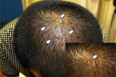
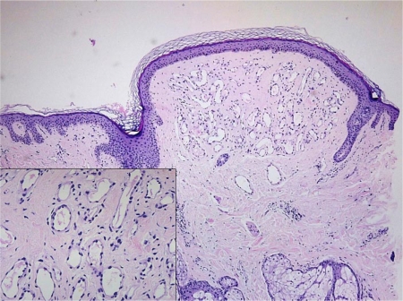
Cherry angiomas are a common cutaneous vascular proliferation which manifests as single or multiple spots and occurs predominantly on the upper trunk and arms. They typically appear as round-to-oval, bright, red, dome-shaped papules and pinpoint macules measuring up to several millimeters in diameter. The histopathologic findings of a cherry angioma are consistent with a true capillary hemangioma, which is formed by numerous, newly developed capillaries with narrow lumens and prominent endothelial cells arranged in a lobular fashion in the papillary dermis. Herein, we report a case of multiple cherry angiomas on the scalp, an uncommon location for cherry angiomas.

摘要

樱桃状血管瘤是一种常见的皮肤血管增生,表现为单个或多个斑点,主要发生在上躯干和手臂。它们通常表现为圆形至椭圆形、明亮的红色、圆顶状丘疹和直径达数毫米的点状斑疹。樱桃状血管瘤的组织病理学表现与真正的毛细血管瘤一致,后者由许多新形成的毛细血管组成,管腔狭窄,内皮细胞突出,以小叶状排列于乳头层真皮内。在此,我们报告一例头皮多发樱桃状血管瘤病例,樱桃状血管瘤出现在头皮这一不常见部位。

https://cdn.ncbi.nlm.nih.gov/pmc/blobs/faaf/2895217/9af17c8a033d/cde0001-0082-f02.jpg
https://cdn.ncbi.nlm.nih.gov/pmc/blobs/faaf/2895217/7f48d73fa3fb/cde0001-0082-f01.jpg
https://cdn.ncbi.nlm.nih.gov/pmc/blobs/faaf/2895217/9af17c8a033d/cde0001-0082-f02.jpg
https://cdn.ncbi.nlm.nih.gov/pmc/blobs/faaf/2895217/7f48d73fa3fb/cde0001-0082-f01.jpg
https://cdn.ncbi.nlm.nih.gov/pmc/blobs/faaf/2895217/9af17c8a033d/cde0001-0082-f02.jpg