• 文献检索
  • 文档翻译
  • 深度研究
  • 学术资讯
  • Suppr Zotero 插件Zotero 插件
  • 邀请有礼
  • 套餐&价格
  • 历史记录
应用&插件
Suppr Zotero 插件Zotero 插件浏览器插件Mac 客户端Windows 客户端微信小程序
定价
高级版会员购买积分包购买API积分包
服务
文献检索文档翻译深度研究API 文档MCP 服务
关于我们
关于 Suppr公司介绍联系我们用户协议隐私条款
关注我们

Suppr 超能文献

核心技术专利:CN118964589B侵权必究
粤ICP备2023148730 号-1Suppr @ 2026

文献检索

告别复杂PubMed语法,用中文像聊天一样搜索,搜遍4000万医学文献。AI智能推荐,让科研检索更轻松。

立即免费搜索

文件翻译

保留排版,准确专业,支持PDF/Word/PPT等文件格式,支持 12+语言互译。

免费翻译文档

深度研究

AI帮你快速写综述,25分钟生成高质量综述,智能提取关键信息,辅助科研写作。

立即免费体验

CdSe/ZnS 量子点对悬浮培养的紫花苜蓿细胞的影响。

The impact of CdSe/ZnS Quantum Dots in cells of Medicago sativa in suspension culture.

机构信息

Biomolecular Diagnostics Laboratory, Instituto de Tecnologia Química e Biológica, Universidade Nova de Lisboa, Apartado 127, 2781-901 Oeiras, Portugal.

出版信息

J Nanobiotechnology. 2010 Oct 7;8:24. doi: 10.1186/1477-3155-8-24.

DOI:10.1186/1477-3155-8-24
PMID:20929583
原文链接:https://pmc.ncbi.nlm.nih.gov/articles/PMC2958997/
Abstract

BACKGROUND

Nanotechnology has the potential to provide agriculture with new tools that may be used in the rapid detection and molecular treatment of diseases and enhancement of plant ability to absorb nutrients, among others. Data on nanoparticle toxicity in plants is largely heterogeneous with a diversity of physicochemical parameters reported, which difficult generalizations. Here a cell biology approach was used to evaluate the impact of Quantum Dots (QDs) nanocrystals on plant cells, including their effect on cell growth, cell viability, oxidative stress and ROS accumulation, besides their cytomobility.

RESULTS

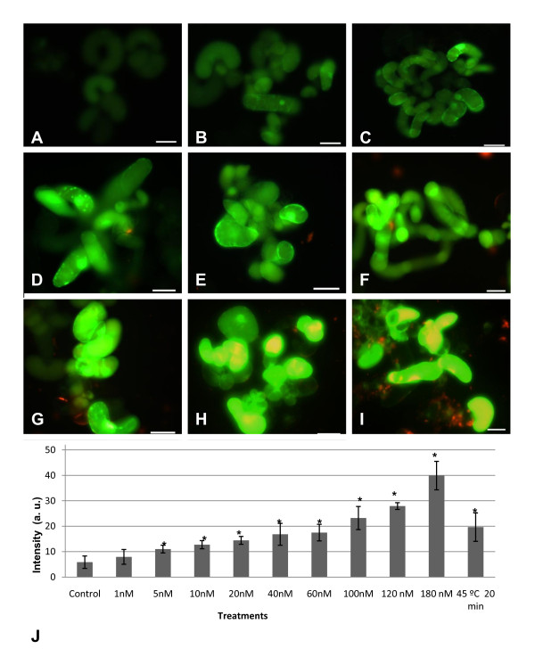
A plant cell suspension culture of Medicago sativa was settled for the assessment of the impact of the addition of mercaptopropanoic acid coated CdSe/ZnS QDs. Cell growth was significantly reduced when 100 mM of mercaptopropanoic acid -QDs was added during the exponential growth phase, with less than 50% of the cells viable 72 hours after mercaptopropanoic acid -QDs addition. They were up taken by Medicago sativa cells and accumulated in the cytoplasm and nucleus as revealed by optical thin confocal imaging. As part of the cellular response to internalization, Medicago sativa cells were found to increase the production of Reactive Oxygen Species (ROS) in a dose and time dependent manner. Using the fluorescent dye H2DCFDA it was observable that mercaptopropanoic acid-QDs concentrations between 5-180 nM led to a progressive and linear increase of ROS accumulation.

CONCLUSIONS

Our results showed that the extent of mercaptopropanoic acid coated CdSe/ZnS QDs cytotoxicity in plant cells is dependent upon a number of factors including QDs properties, dose and the environmental conditions of administration and that, for Medicago sativa cells, a safe range of 1-5 nM should not be exceeded for biological applications.

摘要

背景

纳米技术有可能为农业提供新的工具,可用于快速检测和分子治疗疾病,以及增强植物吸收营养的能力等。关于纳米颗粒对植物的毒性的数据差异很大,报道的理化参数多种多样,难以进行概括。在这里,我们采用细胞生物学方法来评估量子点(QD)纳米晶体对植物细胞的影响,包括它们对细胞生长、细胞活力、氧化应激和 ROS 积累的影响,以及它们的细胞迁移能力。

结果

我们使用紫花苜蓿悬浮细胞培养物来评估添加巯基丙酸(MPA)包覆的 CdSe/ZnS QD 的影响。当在指数生长期添加 100mM 的巯基丙酸-QD 时,细胞生长显著降低,添加巯基丙酸-QD 72 小时后,不到 50%的细胞存活。它们被紫花苜蓿细胞摄取,并在细胞质和核内积累,这一点通过光学薄的共焦成像揭示出来。作为细胞内化反应的一部分,紫花苜蓿细胞被发现以剂量和时间依赖的方式增加活性氧物种(ROS)的产生。使用荧光染料 H2DCFDA,可以观察到 5-180nM 的巯基丙酸-QD 浓度导致 ROS 积累的逐渐和线性增加。

结论

我们的结果表明,MPA 包覆的 CdSe/ZnS QD 在植物细胞中的细胞毒性程度取决于许多因素,包括 QD 的性质、剂量以及给药的环境条件,对于紫花苜蓿细胞,为了进行生物应用,不应超过 1-5nM 的安全范围。

https://cdn.ncbi.nlm.nih.gov/pmc/blobs/a5cd/2958997/3475f1807b1b/1477-3155-8-24-9.jpg

相似文献

1
The impact of CdSe/ZnS Quantum Dots in cells of Medicago sativa in suspension culture.CdSe/ZnS 量子点对悬浮培养的紫花苜蓿细胞的影响。
J Nanobiotechnology. 2010 Oct 7;8:24. doi: 10.1186/1477-3155-8-24.
2
CdSe/ZnS quantum dots trigger DNA repair and antioxidant enzyme systems in Medicago sativa cells in suspension culture.硒化镉/硫化锌量子点在悬浮培养的紫花苜蓿细胞中触发 DNA 修复和抗氧化酶系统。
BMC Biotechnol. 2013 Dec 20;13:111. doi: 10.1186/1472-6750-13-111.
3
Bioaccumulation and toxicity of CdSe/ZnS quantum dots in Phanerochaete chrysosporium.CdSe/ZnS 量子点在黄孢原毛平革菌中的生物积累和毒性。
Colloids Surf B Biointerfaces. 2017 Nov 1;159:303-311. doi: 10.1016/j.colsurfb.2017.08.006. Epub 2017 Aug 5.
4
A Comparison of Common Quantum Dot Alternatives to Cadmium-Based Quantum Dots on the Basis of Liver Cytotoxicity.基于肝细胞毒性对镉基量子点常见替代物的比较
Nanomaterials (Basel). 2024 Jun 25;14(13):1086. doi: 10.3390/nano14131086.
5
CdSe/ZnS quantum dots induce photodynamic effects and cytotoxicity in pancreatic cancer cells.硒化镉/硫化锌量子点诱导胰腺癌细胞产生光动力效应和细胞毒性。
World J Gastroenterol. 2016 Jun 7;22(21):5012-22. doi: 10.3748/wjg.v22.i21.5012.
6
CdSe/ZnS quantum dots exhibited nephrotoxicity through mediating oxidative damage and inflammatory response.硒化镉/硫化锌量子点通过介导氧化损伤和炎症反应表现出肾毒性。
Aging (Albany NY). 2020 Nov 16;13(8):12194-12206. doi: 10.18632/aging.103774.
7
Transcriptome Profile Alteration with Cadmium Selenide/Zinc Sulfide Quantum Dots in .硒化镉/硫化锌量子点对 . 的转录组谱改变
Biomolecules. 2019 Oct 25;9(11):653. doi: 10.3390/biom9110653.
8
Cytotoxicity assessment of functionalized CdSe, CdTe and InP quantum dots in two human cancer cell models.功能化 CdSe、CdTe 和 InP 量子点在两种人类癌细胞模型中的细胞毒性评估。
Mater Sci Eng C Mater Biol Appl. 2015 Dec 1;57:222-31. doi: 10.1016/j.msec.2015.07.044. Epub 2015 Jul 26.
9
Effects of CdSe and CdSe/ZnS Core/Shell Quantum Dots on Singlet Oxygen Production and Cell Toxicity.CdSe和CdSe/ZnS核壳量子点对单线态氧生成及细胞毒性的影响
J Nanosci Nanotechnol. 2018 Mar 1;18(3):1568-1576. doi: 10.1166/jnn.2018.15305.
10
Immunotoxicity assessment of CdSe/ZnS quantum dots in macrophages, lymphocytes and BALB/c mice.CdSe/ZnS量子点在巨噬细胞、淋巴细胞和BALB/c小鼠中的免疫毒性评估
J Nanobiotechnology. 2016 Feb 4;14:10. doi: 10.1186/s12951-016-0162-4.

引用本文的文献

1
Enhancing lettuce yield via Cu/Fe-layered double hydroxide nanoparticles spraying.通过喷洒 Cu/Fe 层状双氢氧化物纳米粒子来提高生菜产量。
J Nanobiotechnology. 2023 Nov 11;21(1):417. doi: 10.1186/s12951-023-02178-6.
2
Sulfur deficiency mediated visible emission of ZnS QDs by magnesium dopant and their application in waste water treatment.硫缺乏通过镁掺杂剂介导硫化锌量子点的可见发射及其在废水处理中的应用。
Heliyon. 2023 Jul 11;9(7):e17947. doi: 10.1016/j.heliyon.2023.e17947. eCollection 2023 Jul.
3
Advantage of Nanotechnology-Based Genome Editing System and Its Application in Crop Improvement.

本文引用的文献

1
Labelling of cells with quantum dots.用量子点标记细胞。
Nanotechnology. 2005 Feb;16(2):R9-R25. doi: 10.1088/0957-4484/16/2/R01. Epub 2005 Jan 25.
2
Experimental approach for an in vitro toxicity assay with non-aggregated quantum dots.使用非聚集量子点进行体外毒性测定的实验方法。
Toxicol In Vitro. 2009 Aug;23(5):955-62. doi: 10.1016/j.tiv.2009.05.007. Epub 2009 May 22.
3
In vitro assessments of nanomaterial toxicity.纳米材料毒性的体外评估。
基于纳米技术的基因组编辑系统的优势及其在作物改良中的应用。
Front Plant Sci. 2021 May 28;12:663849. doi: 10.3389/fpls.2021.663849. eCollection 2021.
4
Nitric oxide and ROS mediate autophagy and regulate Alternaria alternata toxin-induced cell death in tobacco BY-2 cells.一氧化氮和活性氧介导自噬,并调节烟草原生质体 BY-2 细胞中Alternaria alternata 毒素诱导的细胞死亡。
Sci Rep. 2019 Jun 20;9(1):8973. doi: 10.1038/s41598-019-45470-y.
5
Nanotechnology in Plant Science: To Make a Long Story Short.植物科学中的纳米技术:简而言之。
Front Bioeng Biotechnol. 2019 May 29;7:120. doi: 10.3389/fbioe.2019.00120. eCollection 2019.
6
Mycorrhizal Fungi Respond to Resource Inequality by Moving Phosphorus from Rich to Poor Patches across Networks.菌根真菌通过在网络中从富斑块向贫斑块转移磷来响应资源不平等。
Curr Biol. 2019 Jun 17;29(12):2043-2050.e8. doi: 10.1016/j.cub.2019.04.061. Epub 2019 Jun 6.
7
Biochemical synthesis of gold nanoparticles from leaf protein of L. xanthi and their physiological, developmental, and ROS scavenging responses on tobacco plant under stress conditions.从 L. xanthi 的叶蛋白中生物化学合成金纳米粒子及其在胁迫条件下对烟草植株的生理、发育和 ROS 清除反应。
IET Nanobiotechnol. 2019 Feb;13(1):23-29. doi: 10.1049/iet-nbt.2018.5148.
8
Evaluating the Toxic Impacts of Cadmium Selenide Nanoparticles on the Aquatic Plant Lemna minor.评估碲化镉纳米颗粒对水生植物浮萍的毒性影响。
Molecules. 2019 Jan 23;24(3):410. doi: 10.3390/molecules24030410.
9
Flavonoids and darkness lower PCD in senescing Vitis vinifera suspension cell cultures.黄酮类化合物和黑暗条件可降低衰老的葡萄悬浮细胞培养物中的程序性细胞死亡。
BMC Plant Biol. 2016 Oct 26;16(1):233. doi: 10.1186/s12870-016-0917-y.
10
Meta-analysis of cellular toxicity for cadmium-containing quantum dots.含镉量子点的细胞毒性的荟萃分析。
Nat Nanotechnol. 2016 May;11(5):479-86. doi: 10.1038/nnano.2015.338. Epub 2016 Feb 29.
Adv Drug Deliv Rev. 2009 Jun 21;61(6):438-56. doi: 10.1016/j.addr.2009.03.005. Epub 2009 Apr 19.
4
Quantum dot weathering results in microbial toxicity.量子点老化会导致微生物毒性。
Environ Sci Technol. 2008 Dec 15;42(24):9424-30. doi: 10.1021/es8023385.
5
Chemical stability of metallic nanoparticles: a parameter controlling their potential cellular toxicity in vitro.金属纳米颗粒的化学稳定性:一个控制其体外潜在细胞毒性的参数。
Environ Pollut. 2009 Apr;157(4):1127-33. doi: 10.1016/j.envpol.2008.10.002. Epub 2008 Nov 14.
6
Fluorescent probes for super-resolution imaging in living cells.用于活细胞超分辨率成像的荧光探针。
Nat Rev Mol Cell Biol. 2008 Dec;9(12):929-43. doi: 10.1038/nrm2531. Epub 2008 Nov 12.
7
Toxicity assessment of manufactured nanomaterials using the unicellular green alga Chlamydomonas reinhardtii.使用单细胞绿藻莱茵衣藻对人造纳米材料进行毒性评估。
Chemosphere. 2008 Oct;73(7):1121-8. doi: 10.1016/j.chemosphere.2008.07.040. Epub 2008 Sep 2.
8
Uptake, translocation, and accumulation of manufactured iron oxide nanoparticles by pumpkin plants.南瓜植株对人造氧化铁纳米颗粒的吸收、转运及积累
J Environ Monit. 2008 Jun;10(6):713-7. doi: 10.1039/b805998e. Epub 2008 May 13.
9
Nanotoxicity: the growing need for in vivo study.纳米毒性:体内研究的迫切需求。
Curr Opin Biotechnol. 2007 Dec;18(6):565-71. doi: 10.1016/j.copbio.2007.11.008. Epub 2007 Dec 21.
10
Measurement of reactive oxygen species in cells and mitochondria.细胞和线粒体中活性氧的测量。
Methods Cell Biol. 2007;80:355-77. doi: 10.1016/S0091-679X(06)80018-X.