Department of Cellular and Molecular Physiology, Institute of Translational Medicine, University of Liverpool, UK.
Cell Calcium. 2011 Jan;49(1):66-77. doi: 10.1016/j.ceca.2010.11.010. Epub 2010 Dec 19.
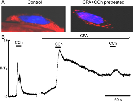
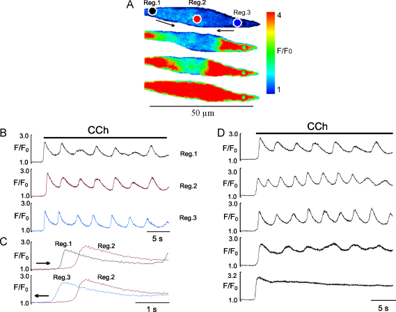
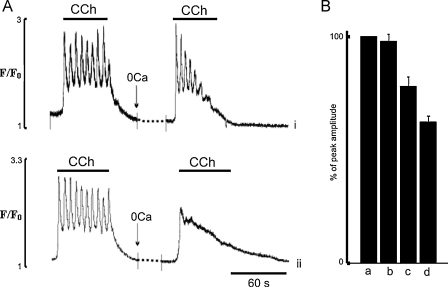
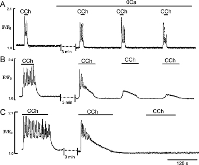
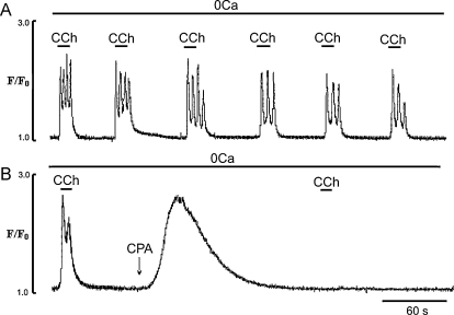
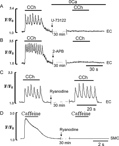
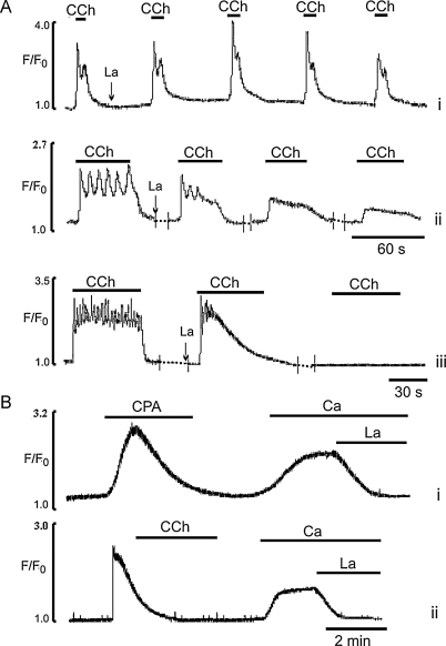
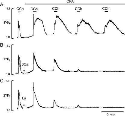
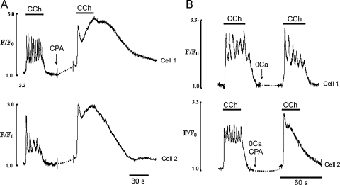
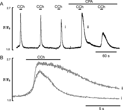
In endothelial cells there remain uncertainties in the details of how Ca(2+) signals are generated and maintained, especially in intact preparations. In particular the role of the sarco-endoplasmic reticulum Ca(2+)-ATPase (SERCA), in contributing to the components of agonist-induced signals is unclear. The aim of this work was to increase understanding of the detailed mechanism of Ca(2+) signalling in endothelial cells using real time confocal imaging of Fluo-4 loaded intact rat tail arteries in response to muscarinic stimulation. In particular we have focused on the role of SERCA, and its interplay with capacitative Ca(2+) entry (CCE) and ER Ca(2+) release and uptake. We have determined its contribution to the Ca(2+) signal and how it varies with different physiological stimuli, including single and repeated carbachol applications and brief and prolonged exposures. In agreement with previous work, carbachol stimulated a rise in intracellular Ca(2+) in the endothelial cells, consisting of a rapid initial phase, then a plateau upon which oscillations of Ca(2+) were superimposed, followed by a decline to basal Ca(2+) levels upon carbachol removal. Our data support the following conclusions: (i) the size (amplitude and duration) of the Ca(2+) spike and early oscillations are limited by SERCA activity, thus both are increased if SERCA is inhibited. (ii) SERCA activity is such that brief applications of carbachol do not trigger CCE, presumably because the fall in luminal Ca(2+) is not sufficient to trigger it. However, longer applications sufficient to deplete the ER or even partial SERCA inhibition stimulate CCE. (iii) Ca(2+) entry occurs via STIM-mediated CCE and SERCA contributes to the cessation of CCE. In conclusion our data show how SERCA function is crucial to shaping endothelial cell Ca signals and its dynamic interplay with both CCE and ER Ca releases.
在内皮细胞中,钙离子信号是如何产生和维持的细节仍存在不确定性,尤其是在完整的标本中。特别是肌浆网钙离子 - ATP酶(SERCA)在激动剂诱导信号成分中的作用尚不清楚。这项工作的目的是通过对装载Fluo - 4的完整大鼠尾动脉进行实时共聚焦成像,以响应毒蕈碱刺激,来增进对内皮细胞中钙离子信号详细机制的理解。特别是,我们重点研究了SERCA的作用,以及它与钙池操纵性钙离子内流(CCE)、内质网钙离子释放和摄取之间的相互作用。我们确定了它对钙离子信号的贡献,以及它如何随不同的生理刺激而变化,包括单次和重复应用卡巴胆碱以及短暂和长时间暴露。与之前的工作一致,卡巴胆碱刺激内皮细胞内钙离子升高,包括一个快速的初始阶段,然后是一个平台期,在此平台期上叠加有钙离子振荡,随后在去除卡巴胆碱后下降至基础钙离子水平。我们的数据支持以下结论:(i)钙离子尖峰和早期振荡的大小(幅度和持续时间)受SERCA活性限制,因此如果SERCA受到抑制,两者都会增加。(ii)SERCA的活性使得短暂应用卡巴胆碱不会触发CCE,大概是因为管腔钙离子的下降不足以触发它。然而,足以耗尽内质网的较长时间应用甚至部分SERCA抑制会刺激CCE。(iii)钙离子内流通过STIM介导的CCE发生,并且SERCA有助于CCE的停止。总之,我们的数据表明SERCA功能对于塑造内皮细胞钙离子信号及其与CCE和内质网钙离子释放的动态相互作用至关重要。