Department of Cancer Genetics, Royal College of Surgeons in Ireland, York House, York Street, Dublin 2, Ireland.
BMC Cancer. 2011 Jan 25;11:33. doi: 10.1186/1471-2407-11-33.
Neuroblastoma is a paediatric cancer which originates from precursor cells of the sympathetic nervous system and accounts for 15% of childhood cancer mortalities. With regards to the role of miRNAs in neuroblastoma, miR-34a, mapping to a chromosome 1p36 region that is commonly deleted, has been found to act as a tumor suppressor through targeting of numerous genes associated with cell proliferation and apoptosis.
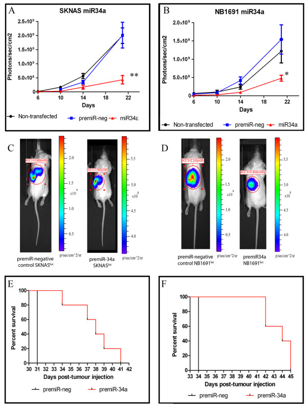
A synthetic miR-34a (or negative control) precursor molecule was transfected into NB1691luc and SK-N-ASluc neuroblastoma cells. Quantitative PCR was used to verify increased miR-34a levels in NB1691luc and SK-N-ASluc cell lines prior to in vitro and in vivo analysis. In vitro analysis of the effects of miR-34a over expression on cell growth, cell cycle and phosphoprotein activation in signal transduction pathways was performed. Neuroblastoma cells over expressing miR-34a were injected retroperitoneally into immunocompromised CB17-SCID mice and tumor burden was assessed over a 21 day period by measuring bioluminescence (photons/sec/cm²).
Over expression of miR-34a in both NB1691luc and SK-N-ASluc neuroblastoma cell lines led to a significant decrease in cell number relative to premiR-negative control treated cells over a 72 hour period. Flow cytometry results indicated that miR-34a induced cell cycle arrest and subsequent apoptosis activation. Phosphoprotein analysis highlighted key elements involved in signal transduction, whose activation was dysregulated as a result of miR-34a introduction into cells. As a potential mechanism of miR-34a action on phosphoprotein levels, we demonstrate that miR-34a over-expression results in a significant reduction of MAP3K9 mRNA and protein levels. Although MAP3K9 is a predicted target of miR-34a, direct targeting could not be validated with luciferase reporter assays. Despite this fact, any functional effects of reduced MAP3K9 expression as a result of miR-34a would be expected to be similar regardless of the mechanism involved. Most notably, in vivo studies showed that tumor growth was significantly repressed after exogenous miR-34a administration in retroperitoneal neuroblastoma tumors.
We demonstrate for the first time that miR-34a significantly reduces tumor growth in an in vivo orthotopic murine model of neuroblastoma and identified novel effects that miR-34a has on phospho-activation of key proteins involved with apoptosis.
神经母细胞瘤是一种起源于交感神经系统前体细胞的小儿癌症,占儿童癌症死亡率的 15%。在 miRNA 与神经母细胞瘤相关的作用方面,miR-34a 定位于染色体 1p36 区域,该区域通常缺失,已被发现通过靶向与细胞增殖和凋亡相关的众多基因作为肿瘤抑制因子发挥作用。
将合成的 miR-34a(或阴性对照)前体分子转染到 NB1691luc 和 SK-N-ASluc 神经母细胞瘤细胞中。在进行体外和体内分析之前,使用定量 PCR 验证 NB1691luc 和 SK-N-ASluc 细胞系中 miR-34a 水平的增加。进行体外分析,以研究 miR-34a 过表达对细胞生长、细胞周期和信号转导途径中磷酸化蛋白激活的影响。将过表达 miR-34a 的神经母细胞瘤细胞注入免疫缺陷型 CB17-SCID 小鼠的腹膜后,并通过测量生物发光(光子/秒/平方厘米)在 21 天的时间内评估肿瘤负担。
在 NB1691luc 和 SK-N-ASluc 神经母细胞瘤细胞系中过表达 miR-34a 导致细胞数量在 72 小时内相对于 premiR-阴性对照处理的细胞显著减少。流式细胞术结果表明,miR-34a 诱导细胞周期停滞和随后的细胞凋亡激活。磷酸化蛋白分析突出了信号转导中涉及的关键元件,其激活由于 miR-34a 引入细胞而失调。作为 miR-34a 对磷酸化蛋白水平作用的潜在机制,我们证明 miR-34a 过表达导致 MAP3K9 mRNA 和蛋白水平的显著降低。尽管 MAP3K9 是 miR-34a 的预测靶标,但不能通过荧光素酶报告基因测定验证直接靶向。尽管如此,由于 miR-34a 导致 MAP3K9 表达减少,任何功能影响都应该是相似的,而不管涉及的机制如何。值得注意的是,体内研究表明,在神经母细胞瘤腹膜后肿瘤中给予外源性 miR-34a 后,肿瘤生长明显受到抑制。
我们首次证明 miR-34a 显著降低神经母细胞瘤在体内原位小鼠模型中的肿瘤生长,并确定了 miR-34a 对涉及凋亡的关键磷酸化蛋白激活的新作用。