Department of Molecular Medicine and Surgery, Karolinska Institutet, Karolinska University Hospital, Stockholm, Sweden.
PLoS One. 2011 Apr 5;6(4):e18213. doi: 10.1371/journal.pone.0018213.
Voltage-dependent K(+) channels (Kv) mediate repolarisation of β-cell action potentials, and thereby abrogate insulin secretion. The role of the Kv1.1 K(+) channel in this process is however unclear. We tested for presence of Kv1.1 in different species and tested for a functional role of Kv1.1 by assessing pancreatic islet function in BALB/cByJ (wild-type) and megencephaly (mceph/mceph) mice, the latter having a deletion in the Kv1.1 gene.
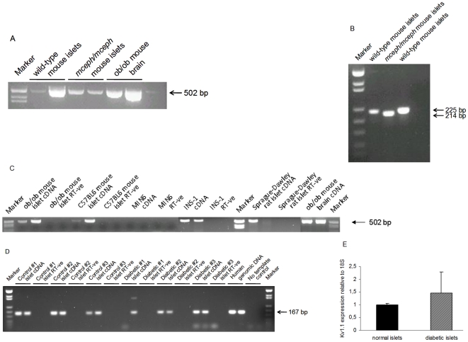
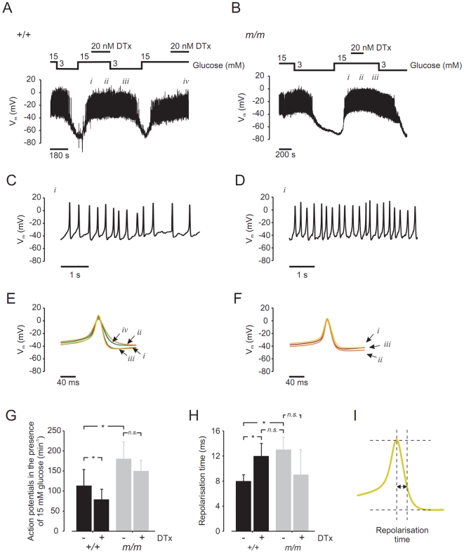
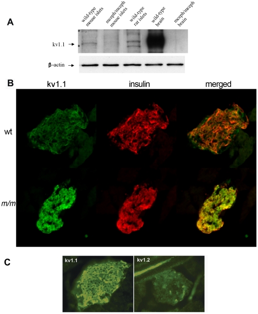
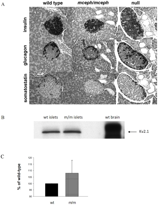
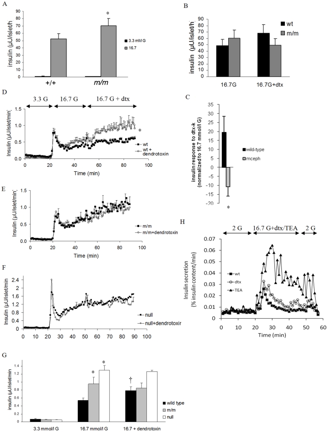
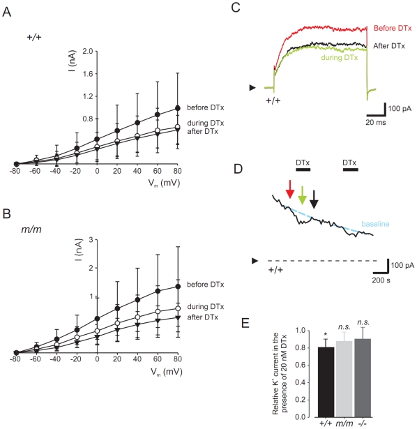
METHODOLOGY/PRINCIPAL FINDINGS: Kv1.1 expression was detected in islets from wild-type mice, SD rats and humans, and expression of truncated Kv1.1 was detected in mceph/mceph islets. Full-length Kv1.1 protein was present in islets from wild-type mice, but, as expected, not in those from mceph/mceph mice. Kv1.1 expression was localized to the β-cell population and also to α- and δ-cells, with evidence of over-expression of truncated Kv1.1 in mceph/mceph islets. Blood glucose, insulin content, and islet morphology were normal in mceph/mceph mice, but glucose-induced insulin release from batch-incubated islets was (moderately) higher than that from wild-type islets. Reciprocal blocking of Kv1.1 by dendrotoxin-K increased insulin secretion from wild-type but not mceph/mceph islets. Glucose-induced action potential duration, as well as firing frequency, was increased in mceph/mceph mouse β-cells. This duration effect on action potential in β-cells from mceph/mceph mice was mimicked by dendrotoxin-K in β-cells from wild-type mice. Observations concerning the effects of both the mceph mutation, and of dendrotoxin-K, on glucose-induced insulin release were confirmed in pancreatic islets from Kv1.1 null mice.
CONCLUSION/SIGNIFICANCE: Kv1.1 channels are expressed in the β-cells of several species, and these channels can influence glucose-stimulated insulin release.
电压门控钾 (Kv) 通道 (Kv) 介导 β 细胞动作电位的复极化,从而终止胰岛素分泌。然而,Kv1.1 K(+) 通道在此过程中的作用尚不清楚。我们检测了不同物种中 Kv1.1 的存在,并通过评估 BALB/cByJ(野生型)和巨脑症(mceph/mceph)小鼠的胰岛功能来测试 Kv1.1 的功能作用,后者的 Kv1.1 基因缺失。
方法/主要发现:Kv1.1 在野生型小鼠、SD 大鼠和人类的胰岛中均有表达,在 mceph/mceph 胰岛中检测到截断的 Kv1.1 表达。全长 Kv1.1 蛋白存在于野生型小鼠的胰岛中,但正如预期的那样,不存在于 mceph/mceph 小鼠的胰岛中。Kv1.1 表达定位于β细胞群体,也定位于α和δ细胞,并且 mceph/mceph 胰岛中截断 Kv1.1 的过表达证据。mceph/mceph 小鼠的血糖、胰岛素含量和胰岛形态正常,但批孵胰岛中葡萄糖诱导的胰岛素释放(适度)高于野生型胰岛。树突毒素-K 对 Kv1.1 的反向阻断增加了野生型但不是 mceph/mceph 胰岛的胰岛素分泌。mceph/mceph 小鼠β细胞中的葡萄糖诱导动作电位持续时间以及放电频率增加。mceph/mceph 小鼠β细胞中树突毒素-K 对动作电位持续时间的这种影响在野生型小鼠的β细胞中被模拟。mceph 突变和树突毒素-K 对葡萄糖诱导胰岛素释放的影响在 Kv1.1 缺失小鼠的胰岛中得到了证实。
结论/意义:Kv1.1 通道在几种物种的β细胞中表达,这些通道可以影响葡萄糖刺激的胰岛素释放。