• 文献检索
  • 文档翻译
  • 深度研究
  • 学术资讯
  • Suppr Zotero 插件Zotero 插件
  • 邀请有礼
  • 套餐&价格
  • 历史记录
应用&插件
Suppr Zotero 插件Zotero 插件浏览器插件Mac 客户端Windows 客户端微信小程序
定价
高级版会员购买积分包购买API积分包
服务
文献检索文档翻译深度研究API 文档MCP 服务
关于我们
关于 Suppr公司介绍联系我们用户协议隐私条款
关注我们

Suppr 超能文献

核心技术专利:CN118964589B侵权必究
粤ICP备2023148730 号-1Suppr @ 2026

文献检索

告别复杂PubMed语法,用中文像聊天一样搜索,搜遍4000万医学文献。AI智能推荐,让科研检索更轻松。

立即免费搜索

文件翻译

保留排版,准确专业,支持PDF/Word/PPT等文件格式,支持 12+语言互译。

免费翻译文档

深度研究

AI帮你快速写综述,25分钟生成高质量综述,智能提取关键信息,辅助科研写作。

立即免费体验

儿童已知骨肉瘤的 CT 肺部结节特征有助于区分结节的良恶性吗?

Do characteristics of pulmonary nodules on computed tomography in children with known osteosarcoma help distinguish whether the nodules are malignant or benign?

机构信息

Department of Radiology, Memorial Sloan-Kettering Cancer Center, New York, NY 10065, USA; Department of Radiology, Division of General and Pediatric Radiology, Medical University Vienna, Vienna General Hospital, 1090 Wien, Austria.

Department of Radiology, Memorial Sloan-Kettering Cancer Center, New York, NY 10065, USA.

出版信息

J Pediatr Surg. 2011 Apr;46(4):729-735. doi: 10.1016/j.jpedsurg.2010.11.027.

DOI:10.1016/j.jpedsurg.2010.11.027
PMID:21496545
原文链接:https://pmc.ncbi.nlm.nih.gov/articles/PMC4443702/
Abstract

PURPOSE

To determine if selected computed tomography (CT) characteristics of pulmonary nodules in pediatric patients with osteosarcoma can help distinguish the nodules as benign or malignant.

METHODS

The institutional review board approved this HIPAA (Health Insurance Portability and Accountability Act-compliant, retrospective study of 30 pediatric osteosarcoma patients (median age 14 years, range 8-22) who underwent chest CT with resection of 117 pulmonary nodules from January 2001 to December 2006. Two pediatric radiologists and one chest radiologist independently and retrospectively reviewed the CT scans and classified nodules as benign, malignant, or indeterminate on the basis of nodule size, laterality, number, location, growth, density, margin appearance, and calcification. Generalized estimating equations were used to examine which characteristics were independent predictors of nodule malignancy.

RESULTS

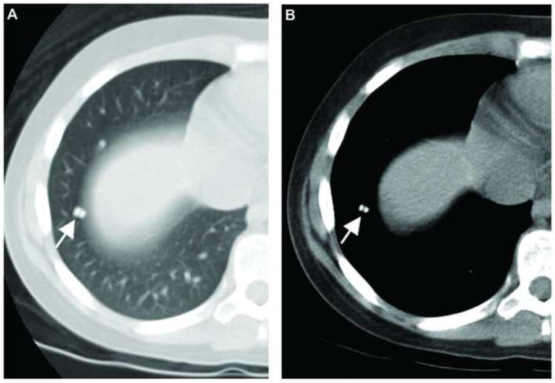
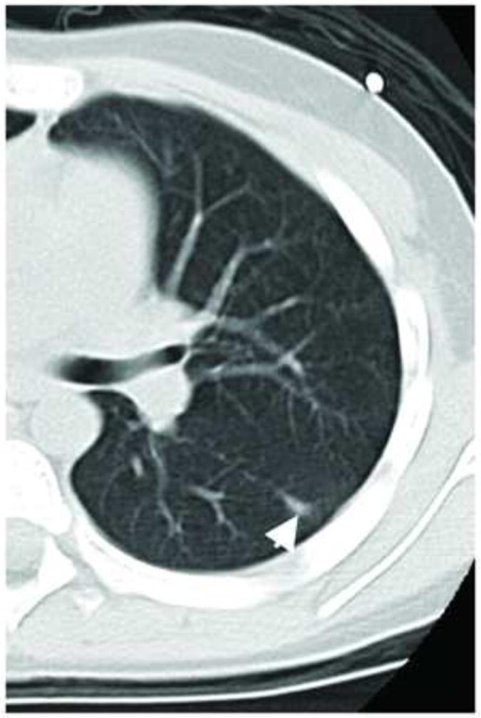
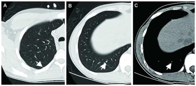
Of the 117 nodules, 80 (68%) were malignant and 37 (32%) were benign by pathologic review. The readers correctly classified 93% to 94% of the malignant nodules. For benign lesions, the results were not as accurate, with the readers correctly classifying only 11% to 30% of lesions. Most of the benign lesions were classified as indeterminate by the readers (54%-65%). Nodule size (≥5 mm) and the presence of calcifications were associated with an increased probability of malignancy (P b .05).

CONCLUSION

On chest CT, nodule size 5 mm or greater and the presence of calcifications are associated with an increased probability of malignant nodule histology in pediatric patients with osteosarcoma. However, nodule characteristics, apart from size and calcification, at chest CT cannot reliably distinguish benign from malignant pulmonary nodules in these patients.

摘要

目的

确定小儿骨肉瘤患者肺部结节的 CT 特征是否有助于区分良恶性。

方法

该机构审查委员会批准了这项 HIPAA(符合健康保险携带和责任法案)合规、回顾性研究,共纳入 30 名小儿骨肉瘤患者(中位年龄 14 岁,范围 8-22 岁),他们于 2001 年 1 月至 2006 年 12 月期间接受胸部 CT 检查并切除了 117 个肺部结节。两名儿科放射科医生和一名胸部放射科医生独立且回顾性地对 CT 扫描进行了评估,并根据结节大小、侧别、数量、位置、生长、密度、边缘外观和钙化情况将结节分为良性、恶性或不确定。广义估计方程用于检查哪些特征是结节恶性的独立预测因素。

结果

117 个结节中,病理检查 80 个(68%)为恶性,37 个(32%)为良性。读者正确分类了 93%-94%的恶性结节。对于良性病变,结果并不准确,读者仅正确分类了 11%-30%的病变。大多数良性病变被读者归类为不确定(54%-65%)。结节大小(≥5mm)和钙化的存在与恶性的可能性增加相关(P b.05)。

结论

在胸部 CT 上,结节大小为 5mm 或更大且存在钙化与小儿骨肉瘤患者恶性结节组织学的可能性增加相关。然而,除了大小和钙化外,这些患者胸部 CT 上的结节特征不能可靠地区分良性和恶性肺部结节。

https://cdn.ncbi.nlm.nih.gov/pmc/blobs/e744/4443702/ea4b7f41033f/nihms460235f3.jpg
https://cdn.ncbi.nlm.nih.gov/pmc/blobs/e744/4443702/7ebc1904d05e/nihms460235f1.jpg
https://cdn.ncbi.nlm.nih.gov/pmc/blobs/e744/4443702/7a1205b24229/nihms460235f2.jpg
https://cdn.ncbi.nlm.nih.gov/pmc/blobs/e744/4443702/ea4b7f41033f/nihms460235f3.jpg
https://cdn.ncbi.nlm.nih.gov/pmc/blobs/e744/4443702/7ebc1904d05e/nihms460235f1.jpg
https://cdn.ncbi.nlm.nih.gov/pmc/blobs/e744/4443702/7a1205b24229/nihms460235f2.jpg
https://cdn.ncbi.nlm.nih.gov/pmc/blobs/e744/4443702/ea4b7f41033f/nihms460235f3.jpg

相似文献

1
Do characteristics of pulmonary nodules on computed tomography in children with known osteosarcoma help distinguish whether the nodules are malignant or benign?儿童已知骨肉瘤的 CT 肺部结节特征有助于区分结节的良恶性吗?
J Pediatr Surg. 2011 Apr;46(4):729-735. doi: 10.1016/j.jpedsurg.2010.11.027.
2
Clinical significance of pulmonary nodules detected on abdominal CT in pediatric patients.小儿腹部CT检查发现肺部结节的临床意义
Pediatr Radiol. 2015 Nov;45(12):1753-60. doi: 10.1007/s00247-015-3407-8. Epub 2015 Jul 12.
3
Distinguishing benign from malignant pulmonary nodules with helical chest CT in children with malignant solid tumors.利用螺旋胸部CT鉴别患有恶性实体瘤儿童的肺结节是良性还是恶性。
Radiology. 2006 May;239(2):514-20. doi: 10.1148/radiol.2392050631.
4
Role of baseline nodule density and changes in density and nodule features in the discrimination between benign and malignant solid indeterminate pulmonary nodules.基线结节密度以及密度和结节特征变化在鉴别实性不确定肺结节良恶性中的作用
Eur J Radiol. 2009 Jun;70(3):492-8. doi: 10.1016/j.ejrad.2008.02.022. Epub 2008 Apr 15.
5
How Are Indeterminate Pulmonary Nodules at Diagnosis Associated with Survival in Patients with High-Grade Osteosarcoma?初诊时的不确定肺部结节与高级别骨肉瘤患者的生存有何关联?
Clin Orthop Relat Res. 2021 Feb 1;479(2):298-308. doi: 10.1097/CORR.0000000000001491.
6
Estimation of malignancy of pulmonary nodules at CT scans: Effect of computer-aided diagnosis on diagnostic performance of radiologists.CT 扫描中肺结节恶性程度的评估:计算机辅助诊断对放射科医生诊断性能的影响。
Asia Pac J Clin Oncol. 2021 Jun;17(3):216-221. doi: 10.1111/ajco.13362. Epub 2020 Aug 5.
7
Limited value of shape, margin and CT density in the discrimination between benign and malignant screen detected solid pulmonary nodules of the NELSON trial.在NELSON试验中,形态、边缘和CT密度在鉴别筛查发现的实性肺结节良恶性方面价值有限。
Eur J Radiol. 2008 Nov;68(2):347-52. doi: 10.1016/j.ejrad.2007.08.027. Epub 2007 Oct 24.
8
Incidental pulmonary nodules detected on abdominal computed tomography.腹部计算机断层扫描发现的偶然肺结节。
J Comput Assist Tomogr. 2012 Nov-Dec;36(6):641-5. doi: 10.1097/RCT.0b013e31826a0de2.
9
Risk of Malignancy in Incidentally Detected Lung Nodules in Patients Aged Younger Than 35 Years.35岁以下患者偶然发现的肺结节的恶性风险
J Comput Assist Tomogr. 2024;48(5):770-773. doi: 10.1097/RCT.0000000000001592. Epub 2024 Feb 27.
10
Thoracoscopic resection of computed tomography-localized lung nodules in children.胸腔镜下切除儿童 CT 定位肺结节。
J Pediatr Surg. 2013 Apr;48(4):750-6. doi: 10.1016/j.jpedsurg.2012.09.051.

引用本文的文献

1
Multimodal Imaging of Osteosarcoma: From First Diagnosis to Radiomics.骨肉瘤的多模态成像:从初次诊断到影像组学
Cancers (Basel). 2025 Feb 10;17(4):599. doi: 10.3390/cancers17040599.
2
Imaging of Osteosarcoma: Presenting Findings, Metastatic Patterns, and Features Related to Prognosis.骨肉瘤的影像学:呈现的表现、转移模式及与预后相关的特征
J Clin Med. 2024 Sep 25;13(19):5710. doi: 10.3390/jcm13195710.
3
Imaging features and clinical evaluation of pulmonary nodules in children.儿童肺结节的影像学特征及临床评估

本文引用的文献

1
Dose-intensified compared with standard chemotherapy for nonmetastatic Ewing sarcoma family of tumors: a Children's Oncology Group Study.与标准化疗相比,剂量强化化疗用于非转移性尤因肉瘤家族性肿瘤:儿童肿瘤学组研究
J Clin Oncol. 2009 May 20;27(15):2536-41. doi: 10.1200/JCO.2008.19.1478. Epub 2009 Apr 6.
2
Soft tissue sarcomas of childhood and adolescence: the prognostic role of tumor size in relation to patient body size.儿童和青少年软组织肉瘤:肿瘤大小与患者体型的关系对预后的影响
J Clin Oncol. 2009 Jan 20;27(3):371-6. doi: 10.1200/JCO.2007.15.4542. Epub 2008 Dec 8.
3
Prognostic factors and long-term results of pulmonary metastasectomy for pediatric histologies.
Front Oncol. 2024 Aug 8;14:1385600. doi: 10.3389/fonc.2024.1385600. eCollection 2024.
4
Predictors of pulmonary metastases on chest computed tomography in children and adolescents with osteosarcoma-tips for qualifying patients for thoracotomy.在儿童和青少年骨肉瘤患者的胸部 CT 上预测肺转移——为开胸手术患者定性的提示。
BMC Pediatr. 2024 Jun 3;24(1):382. doi: 10.1186/s12887-024-04858-0.
5
Deep convolutional neural network based on CT images of pulmonary nodules in the lungs of adolescent and young adult patients with osteosarcoma.基于骨肉瘤青少年及青年成年患者肺部CT图像的深度卷积神经网络。
Oncol Lett. 2023 Jun 23;26(2):344. doi: 10.3892/ol.2023.13930. eCollection 2023 Aug.
6
CT features of osteosarcoma lung metastasis: a retrospective study of 127 patients.CT 特征在骨肉瘤肺转移中的表现:127 例患者的回顾性研究。
J Bras Pneumol. 2023 Apr 28;49(2):e20220433. doi: 10.36416/1806-3756/e20220433. eCollection 2023.
7
The Role of a Multidisciplinary Team in the Diagnosis and Treatment of Bone and Soft Tissue Sarcomas: A Single-Center Experience.多学科团队在骨肉瘤和软组织肉瘤诊断与治疗中的作用:单中心经验
J Pers Med. 2022 Dec 16;12(12):2079. doi: 10.3390/jpm12122079.
8
Osteosarcoma immune prognostic index can indicate the nature of indeterminate pulmonary nodules and predict the metachronous metastasis in osteosarcoma patients.骨肉瘤免疫预后指数可提示骨肉瘤患者中不确定肺结节的性质,并预测异时转移。
Front Oncol. 2022 Jul 22;12:952228. doi: 10.3389/fonc.2022.952228. eCollection 2022.
9
Radiomics Analysis of Multiparametric MRI for Prediction of Synchronous Lung Metastases in Osteosarcoma.多参数MRI的影像组学分析用于预测骨肉瘤的同步肺转移
Front Oncol. 2022 Feb 22;12:802234. doi: 10.3389/fonc.2022.802234. eCollection 2022.
10
The diagnosis of pulmonary metastases on chest computed tomography in primary bone sarcoma and musculoskeletal soft tissue sarcoma.胸部 CT 对原发性骨肉瘤和肌骨软组织肉瘤肺转移的诊断。
Br J Radiol. 2021 Jul 1;94(1123):20210088. doi: 10.1259/bjr.20210088. Epub 2021 May 21.
小儿组织学类型肺转移瘤切除术的预后因素及长期结果
Eur J Cardiothorac Surg. 2008 Dec;34(6):1240-6. doi: 10.1016/j.ejcts.2008.07.039. Epub 2008 Oct 1.
4
Prognostic factors in metastatic rhabdomyosarcomas: results of a pooled analysis from United States and European cooperative groups.转移性横纹肌肉瘤的预后因素:来自美国和欧洲合作组的汇总分析结果
J Clin Oncol. 2008 May 10;26(14):2384-9. doi: 10.1200/JCO.2007.14.7207.
5
Helical computed tomography inaccuracy in the detection of pulmonary metastases: can it be improved?螺旋计算机断层扫描在检测肺转移瘤方面的准确性:能否得到改善?
Ann Thorac Surg. 2007 Dec;84(6):1830-6. doi: 10.1016/j.athoracsur.2007.06.069.
6
Indeterminate pulmonary nodules in patients with sarcoma affect survival.肉瘤患者的肺结节性质不定会影响生存。
Clin Orthop Relat Res. 2007 Jun;459:118-21. doi: 10.1097/BLO.0b013e31805d8606.
7
ACR Appropriateness Criteria on solitary pulmonary nodule.美国放射学会关于孤立性肺结节的适宜性标准
J Am Coll Radiol. 2007 Mar;4(3):152-5. doi: 10.1016/j.jacr.2006.12.003.
8
Distinguishing benign from malignant pulmonary nodules with helical chest CT in children with malignant solid tumors.利用螺旋胸部CT鉴别患有恶性实体瘤儿童的肺结节是良性还是恶性。
Radiology. 2006 May;239(2):514-20. doi: 10.1148/radiol.2392050631.
9
Computed tomographic scan of the chest underestimates the number of metastatic lesions in osteosarcoma.胸部计算机断层扫描会低估骨肉瘤转移灶的数量。
J Pediatr Surg. 2006 Jan;41(1):200-6; discussion 200-6. doi: 10.1016/j.jpedsurg.2005.10.024.
10
Accuracy of helical CT in the detection of pulmonary metastases: is intraoperative palpation still necessary?螺旋CT检测肺转移瘤的准确性:术中触诊是否仍有必要?
Ann Thorac Surg. 2004 Dec;78(6):1910-6; discussion 1916-8. doi: 10.1016/j.athoracsur.2004.05.065.