Department of Clinical Pathology and Immunology, Kobe University Graduate School of Medicine, 7-5-2, Kusunoki-cho, Chuo-ku, Kobe 650-0017, Japan.
Arthritis Res Ther. 2011 May 18;13(3):R77. doi: 10.1186/ar3339.
The purpose of this study was to elucidate the effects of histone deacetylase inhibition on the phenotype and function of dendritic cells and on arthritis in SKG mice.
Arthritis was induced in SKG mice by zymosan A injection. Trichostatin A, a histone deacetylase inhibitor, was administered and its effects on arthritis were evaluated by joint swelling and histological evaluation. Interleukin-17 production in lymph node cells was determined by an enzyme-linked immunosorbent assay (ELISA). Foxp3 expression in lymph node cells and the phenotypes of splenic dendritic cells were examined by fluorescence-activated cell sorting (FACS). Bone marrow-derived dendritic cells (BM-DC) were generated with granulocyte macrophage colony-stimulating factor. The effects of trichostatin A on cell surface molecules, cytokine production, indoleamine 2,3-dioxygenase (IDO) expression and T cell stimulatory capacity were examined by FACS, ELISA, quantitative real-time polymerase chain reaction and Western blot, and the allo-mixed lymphocyte reaction, respectively.
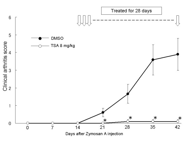
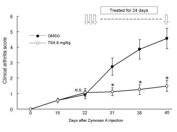
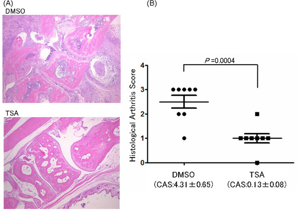
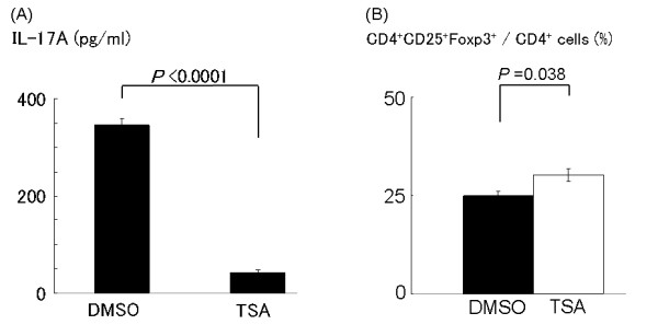
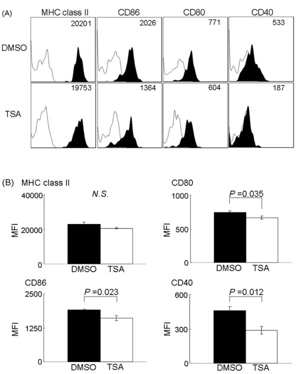
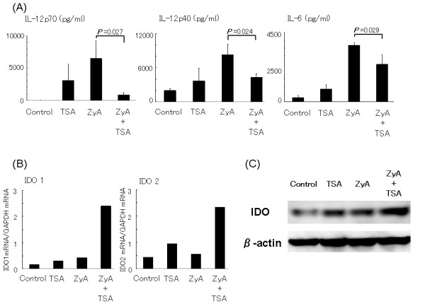
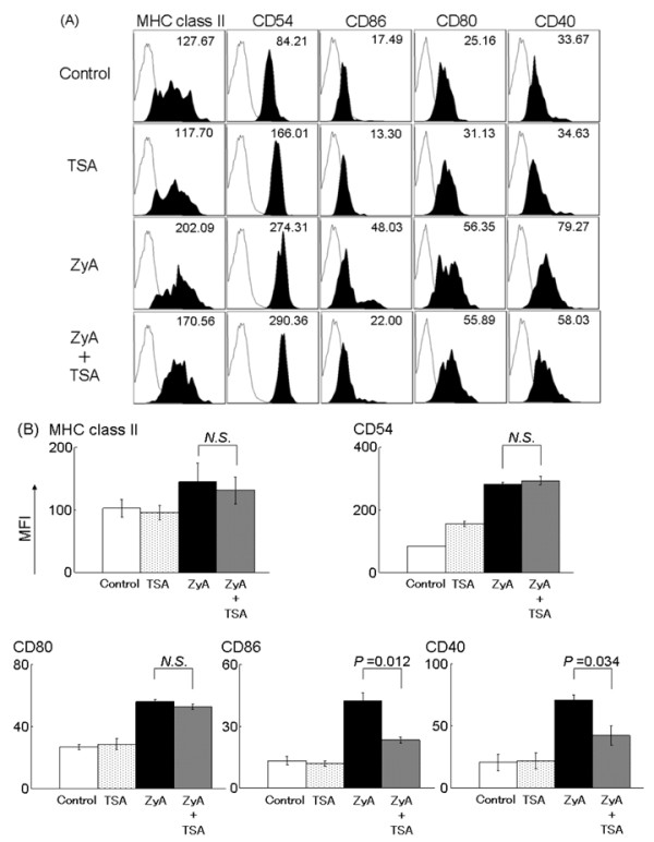
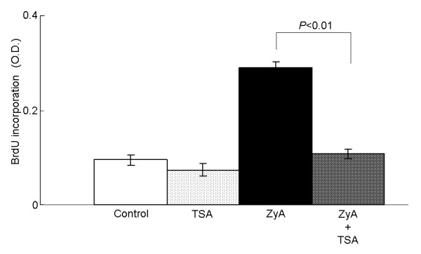
Trichostatin A, when administered before the onset of arthritis, prevented SKG mice from getting arthritis. Trichostatin A treatment also showed therapeutic effects on arthritis in SKG mice, when it was administered after the onset of arthritis. Trichostatin A treatment reduced Th17 cells and induced regulatory T cells in lymph node, and also decreased co-stimulatory molecule expression on splenic dendritic cells in vivo. In vitro, trichostatin A markedly suppressed zymosan A-induced interleukin-12 and interleukin-6 production by BM-DC and up-regulated IDO expression at mRNA and protein levels. Trichostatin A-treated BM-DC also showed less T cell stimulatory capacity.
Histone deacetylase inhibition changes dendritic cells to a tolerogenic phenotype and ameliorates arthritis in SKG mice.
本研究旨在阐明组蛋白去乙酰化酶抑制对树突状细胞表型和功能的影响,以及对 SKG 小鼠关节炎的影响。
通过酵母聚糖 A 注射诱导 SKG 小鼠关节炎。给予组蛋白去乙酰化酶抑制剂曲古抑菌素 A,并通过关节肿胀和组织学评估评估其对关节炎的影响。通过酶联免疫吸附试验(ELISA)测定淋巴结细胞中白细胞介素-17 的产生。通过荧光激活细胞分选(FACS)检测淋巴结细胞中 Foxp3 的表达和脾树突状细胞的表型。用粒细胞巨噬细胞集落刺激因子生成骨髓来源的树突状细胞(BM-DC)。通过 FACS、ELISA、实时定量聚合酶链反应和 Western blot 分别检测曲古抑菌素 A 对细胞表面分子、细胞因子产生、吲哚胺 2,3-双加氧酶(IDO)表达和 T 细胞刺激能力的影响,以及同种混合淋巴细胞反应。
曲古抑菌素 A 在关节炎发病前给药可预防 SKG 小鼠发生关节炎。关节炎发病后给予曲古抑菌素 A 治疗也对 SKG 小鼠关节炎有治疗作用。曲古抑菌素 A 治疗可减少淋巴结中的 Th17 细胞并诱导调节性 T 细胞,并降低体内脾树突状细胞共刺激分子的表达。在体外,曲古抑菌素 A 显著抑制 BM-DC 诱导的白细胞介素-12 和白细胞介素-6 的产生,并上调 IDO 在 mRNA 和蛋白水平的表达。用曲古抑菌素 A 处理的 BM-DC 也显示出较低的 T 细胞刺激能力。
组蛋白去乙酰化酶抑制可使树突状细胞向耐受性表型转变,并改善 SKG 小鼠的关节炎。