Section on Molecular Neurobiology, National Institute of Child Health and Human Development, National Institutes of Health, Bethesda, Maryland 20892, USA.
Biol Psychiatry. 2011 Oct 1;70(7):636-45. doi: 10.1016/j.biopsych.2011.04.016. Epub 2011 Jun 12.
Neuregulin-1 and ErbB4 are genetically associated with schizophrenia, and detailed knowledge of the cellular and subcellular localization of ErbB4 is important for understanding how neuregulin-1 regulates neuronal network activity and behavior. Expression of ErbB4 is restricted to interneurons in the rodent hippocampus and cortex. However, controversy remains about the cellular expression pattern in primate brain and its subcellular distribution in postsynaptic somatodendritic locations versus presynaptic terminals.
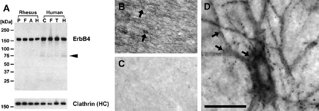
ErbB4 expression was analyzed in pyramidal cells and interneurons in the frontal cortex of five species: C57BL6 mice (n = 3), ErbB4⁻/⁻ mice (n = 2), Sprague-Dawley rats (n = 3), two macaque species (n = 3 + 2), and humans (normal control subjects, n = 2). We investigated 1) messenger RNA in mice, macaques, and humans; 2) protein expression in all species using highly specific monoclonal antibodies; and 3) specificity tests of several ErbB4 antibodies on brain samples (mouse, macaque, human).
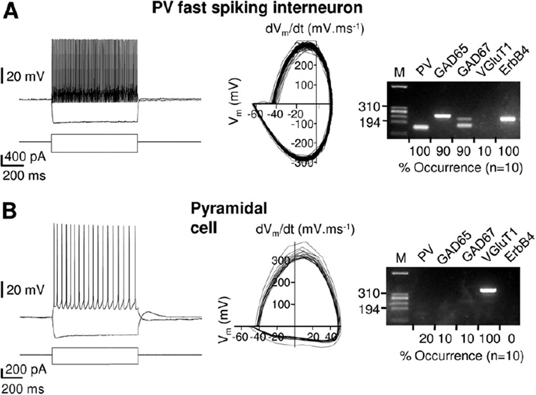
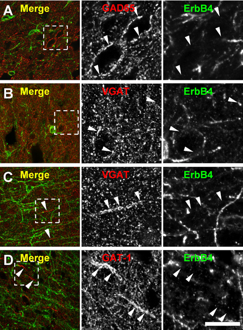
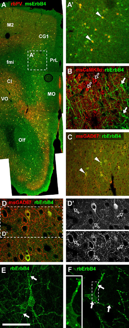
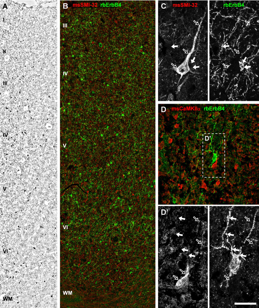
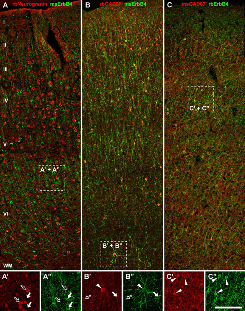
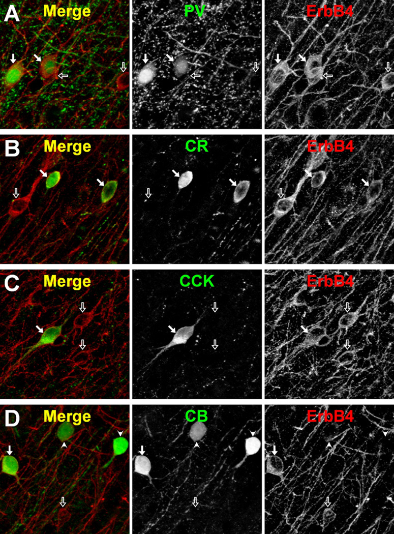
ErbB4 RNA is restricted to interneurons in the frontal cortex of mice. ErbB4 protein is undetectable in pyramidal cells of rodents, macaques, and human frontal cortex, whereas most interneurons positive for parvalbumin, calretinin, or cholecystokinin, but only a minority of calbindin-positive cells, co-express ErbB4 in macaques. Importantly, no presynaptic ErbB4 expression was detected in any species.
The interneuron-selective somatodendritic expression of ErbB4 is consistent with a primary role of neuregulin-ErbB4 signaling in the postsynaptic modulation of gamma-aminobutyric acidergic function in rodents and primates. Our data validate the use of rodents to analyze effects of abnormal ErbB4 function as a means to model endophenotypes of psychiatric disorders.
神经调节蛋白-1 和 ErbB4 与精神分裂症在基因上相关联,详细了解 ErbB4 的细胞和亚细胞定位对于理解神经调节蛋白-1 如何调节神经元网络活动和行为至关重要。ErbB4 的表达仅限于啮齿动物海马体和皮质中的中间神经元。然而,关于灵长类动物大脑中的细胞表达模式及其在突触后体树突部位与突触前末端的亚细胞分布,仍存在争议。
在五种物种的额皮质中的锥体神经元和中间神经元中分析 ErbB4 的表达:C57BL6 小鼠(n = 3)、ErbB4⁻/⁻ 小鼠(n = 2)、斯普拉格-道利大鼠(n = 3)、两种猕猴物种(n = 3 + 2)和人类(正常对照受试者,n = 2)。我们研究了 1)在小鼠、猕猴和人类中的信使 RNA;2)在所有物种中使用高度特异性单克隆抗体进行的蛋白表达;3)几种 ErbB4 抗体在脑组织样本(小鼠、猕猴、人类)上的特异性测试。
ErbB4 RNA 仅限于小鼠额皮质中的中间神经元。在啮齿动物、猕猴和人类额皮质的锥体神经元中,无法检测到 ErbB4 蛋白,而大多数中间神经元对 parvalbumin、calretinin 或 cholecystokinin 呈阳性反应,但只有少数 calbindin 阳性细胞对 ErbB4 呈阳性反应,在猕猴中,ErbB4 共表达。重要的是,在任何物种中均未检测到突触前 ErbB4 表达。
ErbB4 的中间神经元选择性体树突表达与神经调节蛋白-ErbB4 信号在γ-氨基丁酸能功能的突触后调节中发挥主要作用一致,在啮齿动物和灵长类动物中。我们的数据验证了使用啮齿动物来分析异常 ErbB4 功能的影响,作为模拟精神疾病表型的一种手段。