Department of Pharmacology, College of Pharmacy, Seoul National University, Seoul, Korea.
J Biomed Sci. 2011 Jul 4;18(1):48. doi: 10.1186/1423-0127-18-48.
At the beginning of neurogenesis, massive brain cell death occurs and more than 50% of cells are eliminated by apoptosis along with neuronal differentiation. However, few studies were conducted so far regarding the regulation of neural progenitor cells (NPCs) death during development. Because of the physiological role of cell death during development, aberration of normal apoptotic cell death is detrimental to normal organogenesis.Apoptosis occurs in not only neuron but also in NPCs and neuroblast. When growth and survival signals such as EGF or LIF are removed, apoptosis is activated as well as the induction of differentiation. To investigate the regulation of cell death during developmental stage, it is essential to investigate the regulation of apoptosis of NPCs.
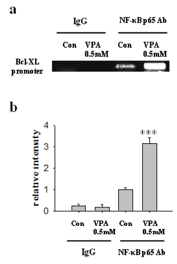
Neural progenitor cells were cultured from E14 embryonic brains of Sprague-Dawley rats. For in vivo VPA animal model, pregnant rats were treated with VPA (400 mg/kg S.C.) diluted with normal saline at E12. To analyze the cell death, we performed PI staining and PARP and caspase-3 cleavage assay. Expression level of proteins was investigated by Western blot and immunocytochemical assays. The level of mRNA expression was investigated by RT-PCR. Interaction of Bcl-XL gene promoter and NF-κB p65 was investigated by ChIP assay.
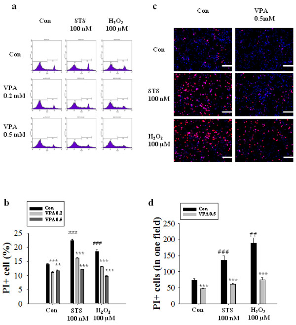
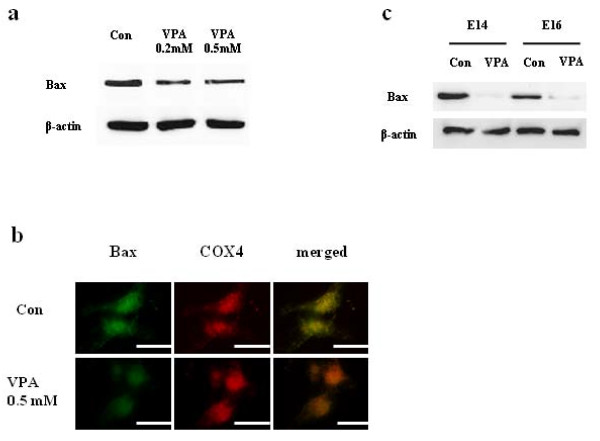
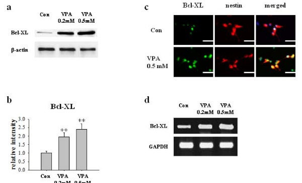
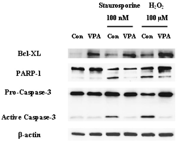
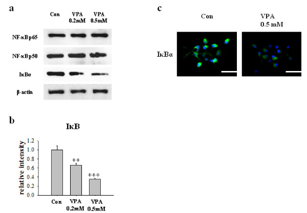
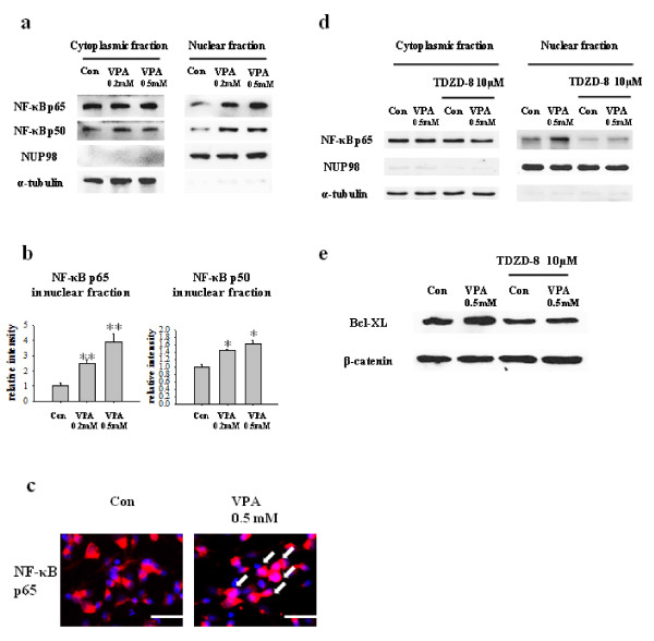
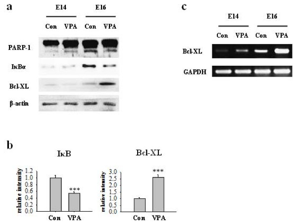
In this study, FACS analysis, PI staining and PARP and caspase-3 cleavage assay showed that VPA protects cultured NPCs from cell death after growth factor withdrawal both in basal and staurosporine- or hydrogen peroxide-stimulated conditions. The protective effect of prenatally injected VPA was also observed in E16 embryonic brain. Treatment of VPA decreased the level of IκBα and increased the nuclear translocation of NF-κB, which subsequently enhanced expression of anti-apoptotic protein Bcl-XL.
To the best of our knowledge, this is the first report to indicate the reduced death of NPCs by VPA at developmentally critical periods through the degradation of IκBα and the activation of NF-κB signaling. The reduced NPCs death might underlie the neurodevelopmental defects collectively called fetal valproate syndrome, which shows symptoms such as mental retardation and autism-like behavior.
在神经发生的早期,大量脑细胞死亡,超过 50%的细胞通过细胞凋亡与神经元分化一起被消除。然而,到目前为止,关于神经祖细胞(NPC)在发育过程中死亡的调节,很少有研究。由于细胞死亡在发育过程中的生理作用,正常凋亡细胞死亡的异常对正常器官发生是有害的。细胞凋亡不仅发生在神经元中,也发生在 NPC 和神经母细胞中。当 EGF 或 LIF 等生长和存活信号被去除时,凋亡被激活,同时诱导分化。为了研究发育阶段细胞死亡的调节,研究 NPC 凋亡的调节是至关重要的。
从 Sprague-Dawley 大鼠 E14 胚胎脑中培养神经祖细胞。对于体内 VPA 动物模型,将怀孕的大鼠用 VPA(400mg/kg S.C.)处理,VPA 用生理盐水稀释,在 E12 时处理。为了分析细胞死亡,我们进行了 PI 染色和 PARP 和 caspase-3 切割测定。通过 Western blot 和免疫细胞化学检测蛋白质的表达水平。通过 RT-PCR 检测 mRNA 表达水平。通过 ChIP 检测 Bcl-XL 基因启动子和 NF-κB p65 的相互作用。
在这项研究中,FACS 分析、PI 染色、PARP 和 caspase-3 切割测定表明,VPA 在基础条件下以及在 staurosporine 或过氧化氢刺激条件下,均可保护培养的 NPC 在生长因子撤出后免于死亡。在 E16 胚胎脑中也观察到了产前注射 VPA 的保护作用。VPA 的处理降低了 IκBα 的水平,并增加了 NF-κB 的核转位,从而增强了抗凋亡蛋白 Bcl-XL 的表达。
据我们所知,这是第一项表明 VPA 通过降解 IκBα 和激活 NF-κB 信号,减少发育关键期 NPC 死亡的报告。NPC 死亡的减少可能是导致被称为胎儿丙戊酸综合征的神经发育缺陷的原因之一,该综合征表现为智力迟钝和自闭症样行为等症状。