Department of Translational Hematology and Oncology Research, Taussig Cancer Institute, Cleveland Clinic, Cleveland, OH 44195, USA.
Leukemia. 2012 Feb;26(2):244-54. doi: 10.1038/leu.2011.207. Epub 2011 Aug 12.
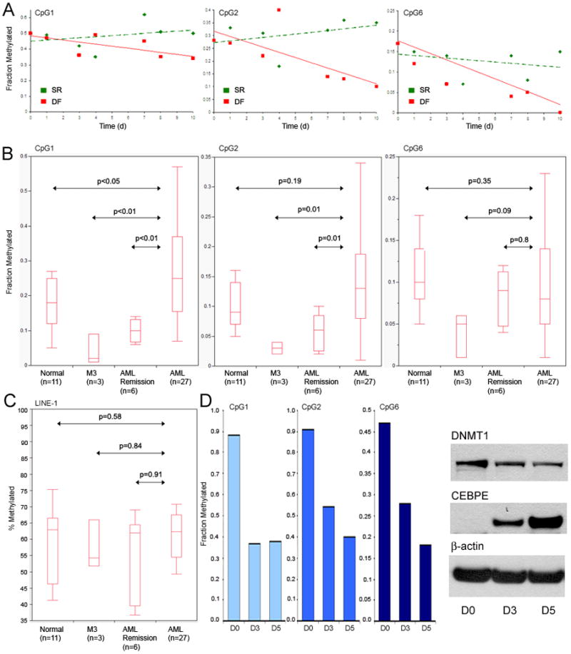
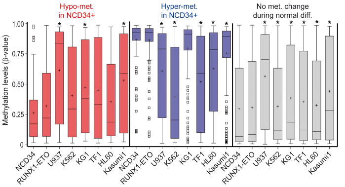
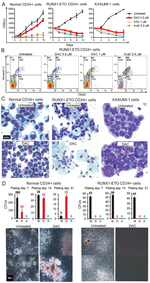
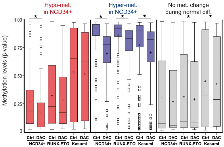
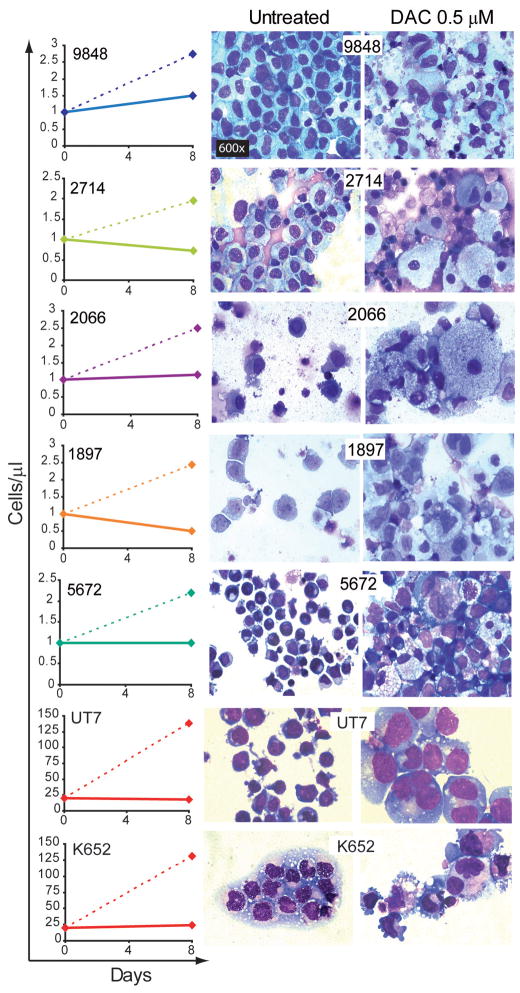
The DNA hypomethylating drug decitabine maintains normal hematopoietic stem cell (HSC) self-renewal but induces terminal differentiation in acute myeloid leukemia (AML) cells. The basis for these contrasting cell fates, and for selective CpG hypomethylation by decitabine, is poorly understood. Promoter CpGs, with methylation measured by microarray, were classified by the direction of methylation change with normal myeloid maturation. In AML cells, the methylation pattern at maturation-responsive CpGs suggested at least partial maturation. Consistent with partial maturation, in gene expression analyses, AML cells expressed high levels of the key lineage-specifying factor CEBPA, but relatively low levels of the key late-differentiation driver CEBPE. In methylation analysis by mass spectrometry, CEBPE promoter CpGs that are usually hypomethylated during granulocyte maturation were significantly hypermethylated in AML cells. Decitabine-induced hypomethylation was greatest at these and other promoter CpGs that are usually hypomethylated with myeloid maturation, accompanied by cellular differentiation of AML cells. In contrast, decitabine-treated normal HSCs retained immature morphology, and methylation significantly decreased at CpGs that are less methylated in immature cells. High expression of lineage-specifying factor and aberrant epigenetic repression of some key late-differentiation driver genes distinguishes AML cells from normal HSCs, and could explain the contrasting differentiation and methylation responses to decitabine.
DNA 去甲基化药物地西他滨维持正常造血干细胞(HSC)的自我更新,但诱导急性髓系白血病(AML)细胞的终末分化。这些相反的细胞命运以及地西他滨对 CpG 的选择性去甲基化的基础尚未完全了解。通过微阵列测量的启动子 CpG 按正常髓样成熟时的甲基化变化方向进行分类。在 AML 细胞中,成熟反应性 CpG 的甲基化模式表明至少部分成熟。与部分成熟一致,在基因表达分析中,AML 细胞表达高水平的关键谱系特异性因子 CEBPA,但相对低水平的关键晚期分化驱动因子 CEBPE。在通过质谱进行的甲基化分析中,在粒细胞成熟过程中通常低甲基化的 CEBPE 启动子 CpG 在 AML 细胞中显著超甲基化。地西他滨诱导的去甲基化在这些和其他在髓样成熟过程中通常低甲基化的启动子 CpG 上最大,伴随着 AML 细胞的细胞分化。相比之下,用地西他滨处理的正常 HSC 保持未成熟的形态,并且在幼稚细胞中甲基化程度较低的 CpG 上甲基化显著降低。谱系特异性因子的高表达和某些关键晚期分化驱动基因的异常表观遗传抑制将 AML 细胞与正常 HSC 区分开来,并可以解释对地西他滨的分化和甲基化反应的差异。