• 文献检索
  • 文档翻译
  • 深度研究
  • 学术资讯
  • Suppr Zotero 插件Zotero 插件
  • 邀请有礼
  • 套餐&价格
  • 历史记录
应用&插件
Suppr Zotero 插件Zotero 插件浏览器插件Mac 客户端Windows 客户端微信小程序
定价
高级版会员购买积分包购买API积分包
服务
文献检索文档翻译深度研究API 文档MCP 服务
关于我们
关于 Suppr公司介绍联系我们用户协议隐私条款
关注我们

Suppr 超能文献

核心技术专利:CN118964589B侵权必究
粤ICP备2023148730 号-1Suppr @ 2026

文献检索

告别复杂PubMed语法,用中文像聊天一样搜索,搜遍4000万医学文献。AI智能推荐,让科研检索更轻松。

立即免费搜索

文件翻译

保留排版,准确专业,支持PDF/Word/PPT等文件格式,支持 12+语言互译。

免费翻译文档

深度研究

AI帮你快速写综述,25分钟生成高质量综述,智能提取关键信息,辅助科研写作。

立即免费体验

儿童脑磁共振成像中多个T2高信号在1型神经纤维瘤病诊断中的临床敏感性和特异性:诊断准确性研究

Clinical sensitivity and specificity of multiple T2-hyperintensities on brain magnetic resonance imaging in diagnosis of neurofibromatosis type 1 in children: diagnostic accuracy study.

作者信息

Sabol Zlatko, Resić Biserka, Gjergja Juraski Romana, Sabol Filip, Kovac Sizgorić Matilda, Orsolić Kresimir, Ozretić David, Sepić-Grahovac Dubravka

机构信息

Sabol Outpatient Clinic for Sick Children, Zagreb, Croatia.

出版信息

Croat Med J. 2011 Aug 15;52(4):488-96. doi: 10.3325/cmj.2011.52.488.

DOI:10.3325/cmj.2011.52.488
PMID:21853543
原文链接:https://pmc.ncbi.nlm.nih.gov/articles/PMC3160695/
Abstract

AIM

To determine the prevalence, number, and location of multiple (≥2) T2-hyperintensities on brain magnetic resonance imaging (MRI) in children with neurofibromatosis type 1 (NF1) and their correlation with age, and to establish their sensitivity, specificity, and accuracy for the diagnosis of NF1 in children, especially in the early age (2-7 years).

METHODS

We performed a cross-sectional study of 162 patients with NF1 from Croatian Neurofibromatosis Association Database and 163 control children between the ages of 2 and 18 years who underwent brain MRI between 1989 and 2009.

RESULTS

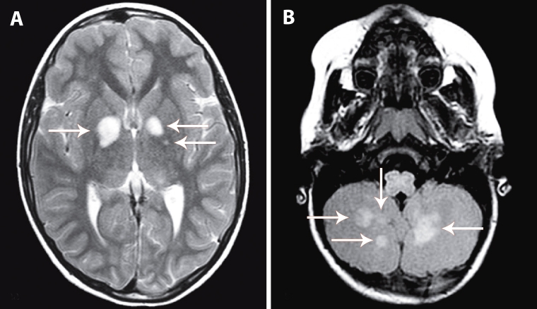
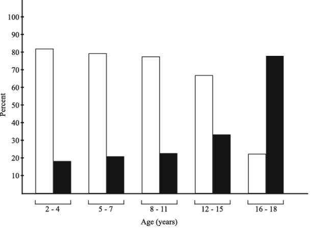
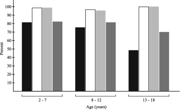
Multiple T2-hyperintensities were present in 74% of NF1 patients and 1.8% of controls. They were mainly located in the basal ganglia, brainstem, and cerebellum and were significantly decreased in prevalence and number in the older age. T2-hyperintensities had excellent diagnostic accuracy with the area under the receiver operating characteristic (ROC) curve of 0.849 and 95% confidence interval (CI) of 0.805-0.886. The diagnostic sensitivity, specificity, and accuracy rate of T2-hyperintensities for NF1 were highest in the youngest age (2-7 years): 81% (95% CI 71%-89.1%), 99% (95% CI 92.3%-100%), and 85.8 (95% CI 83.3-93.8), respectively.

CONCLUSION

This study strongly suggests the inclusion of T2-hyperintensities on brain MRI on the list of diagnostic criteria for NF1, especially in children of early age, when the clinical penetration of the NF1 gene has not yet been completely finished.

摘要

目的

确定1型神经纤维瘤病(NF1)患儿脑磁共振成像(MRI)上多发(≥2个)T2高信号的患病率、数量和位置及其与年龄的相关性,并确定其对儿童尤其是低龄(2 - 7岁)儿童NF1诊断的敏感性、特异性和准确性。

方法

我们对来自克罗地亚神经纤维瘤病协会数据库的162例NF1患者以及163例年龄在2至18岁之间、于1989年至2009年间接受脑MRI检查的对照儿童进行了横断面研究。

结果

74%的NF1患者存在多发T2高信号,而对照儿童中这一比例为1.8%。它们主要位于基底神经节、脑干和小脑,且患病率和数量在大龄儿童中显著降低。T2高信号具有出色的诊断准确性,受试者操作特征(ROC)曲线下面积为0.849,95%置信区间(CI)为0.805 - 0.886。T2高信号对NF1的诊断敏感性、特异性和准确率在最年幼年龄组(2 - 7岁)最高,分别为81%(95%CI 71% - 89.1%)、99%(95%CI 92.3% - 100%)和85.8(95%CI 83.3 - 93.8)。

结论

本研究强烈建议将脑MRI上的T2高信号纳入NF1的诊断标准清单,尤其是在NF1基因的临床外显尚未完全完成的低龄儿童中。

https://cdn.ncbi.nlm.nih.gov/pmc/blobs/4235/3160695/12cacc3d197b/CroatMedJ_52_0488-F5.jpg
https://cdn.ncbi.nlm.nih.gov/pmc/blobs/4235/3160695/f8a4e408b08c/CroatMedJ_52_0488-F1.jpg
https://cdn.ncbi.nlm.nih.gov/pmc/blobs/4235/3160695/af3412b73a0a/CroatMedJ_52_0488-F2.jpg
https://cdn.ncbi.nlm.nih.gov/pmc/blobs/4235/3160695/9a665e06e639/CroatMedJ_52_0488-F3.jpg
https://cdn.ncbi.nlm.nih.gov/pmc/blobs/4235/3160695/523cd9ed6f82/CroatMedJ_52_0488-F4.jpg
https://cdn.ncbi.nlm.nih.gov/pmc/blobs/4235/3160695/12cacc3d197b/CroatMedJ_52_0488-F5.jpg

相似文献

1
Clinical sensitivity and specificity of multiple T2-hyperintensities on brain magnetic resonance imaging in diagnosis of neurofibromatosis type 1 in children: diagnostic accuracy study.儿童脑磁共振成像中多个T2高信号在1型神经纤维瘤病诊断中的临床敏感性和特异性:诊断准确性研究
Croat Med J. 2011 Aug 15;52(4):488-96. doi: 10.3325/cmj.2011.52.488.
2
Is magnetic resonance spectroscopy capable of detecting metabolic abnormalities in neurofibromatosis type 1 that are not revealed in brain parenchyma of normal appearance?磁共振波谱分析能否检测出1型神经纤维瘤病中未在外观正常的脑实质中显示的代谢异常?
Pediatr Neurol. 2015 Mar;52(3):314-9. doi: 10.1016/j.pediatrneurol.2014.11.014. Epub 2014 Dec 1.
3
Natural history of cognitive deficits and their relationship to MRI T2-hyperintensities in NF1.1型神经纤维瘤病认知缺陷的自然病史及其与MRI T2高信号的关系。
Neurology. 2003 Apr 8;60(7):1139-45. doi: 10.1212/01.wnl.0000055090.78351.c1.
4
Spinal Cord Hyperintensities in Neurofibromatosis Type 1: Are They the Cord Equivalent of Unidentified Bright Objects in the Brain?脊髓神经纤维瘤病 1 型中的脊髓高信号:它们是大脑中未识别的明亮物体在脊髓上的对应物吗?
Pediatr Neurol. 2018 Sep;86:63-65. doi: 10.1016/j.pediatrneurol.2018.07.005. Epub 2018 Jul 20.
5
Use of "unidentified bright objects" on MRI for diagnosis of neurofibromatosis 1 in children.利用磁共振成像(MRI)上的“不明明亮物体”诊断儿童1型神经纤维瘤病。
Neurology. 2000 Apr 25;54(8):1646-51. doi: 10.1212/wnl.54.8.1646.
6
Examining the frontal subcortical brain vulnerability hypothesis in children with neurofibromatosis type 1: Are T2-weighted hyperintensities related to executive dysfunction?研究1型神经纤维瘤病患儿的额叶皮质下脑易损性假说:T2加权高信号与执行功能障碍有关吗?
Neuropsychology. 2015 May;29(3):473-84. doi: 10.1037/neu0000151. Epub 2014 Nov 3.
7
Unidentified bright objects on brain MRI in children as a diagnostic criterion for neurofibromatosis type 1.儿童脑部磁共振成像上不明的明亮物体作为1型神经纤维瘤病的诊断标准。
Pediatr Radiol. 2008 Mar;38(3):305-10. doi: 10.1007/s00247-007-0712-x. Epub 2008 Jan 30.
8
MRI abnormalities in neurofibromatosis type 1 (NF1): a study of men and mice.1型神经纤维瘤病(NF1)的MRI异常:一项针对人类和小鼠的研究
Brain Dev. 1999 Jun;21(4):268-73. doi: 10.1016/s0387-7604(99)00024-8.
9
Longitudinal assessment of cognition and T2-hyperintensities in NF1: an 18-year study.NF1 患者认知功能和 T2 高信号的纵向评估:一项 18 年的研究。
Am J Med Genet A. 2014 Mar;164A(3):661-5. doi: 10.1002/ajmg.a.36338. Epub 2013 Dec 19.
10
Brain morphometry, T2-weighted hyperintensities, and IQ in children with neurofibromatosis type 1.1型神经纤维瘤病患儿的脑形态测量、T2加权高信号与智商
Arch Neurol. 2005 Dec;62(12):1904-8. doi: 10.1001/archneur.62.12.1904.

引用本文的文献

1
Neuroimaging Reader Study on Clinical Sensitivity and Specificity Using Synthetic MRI Based on MR Quantification.基于磁共振定量的合成磁共振成像在临床敏感性和特异性方面的神经影像学读者研究
AJNR Am J Neuroradiol. 2025 Jun 3;46(6):1196-1202. doi: 10.3174/ajnr.A8631.
2
Non-Optic Glioma-like Lesions in Adult Neurofibromatosis Type 1 Patients.1型成人神经纤维瘤病患者中的非视神经胶质瘤样病变
Diagnostics (Basel). 2024 Dec 30;15(1):67. doi: 10.3390/diagnostics15010067.
3
The role of visual electrodiagnostics in management of children with neurofibromatosis type 1.

本文引用的文献

1
Thalamo-striatal T2-weighted hyperintensities (unidentified bright objects) correlate with cognitive impairments in neurofibromatosis type 1 during childhood.丘脑纹状体T2加权高信号(不明亮物体)与1型神经纤维瘤病患儿的认知障碍相关。
Dev Neuropsychol. 2009;34(6):736-48. doi: 10.1080/87565640903265137.
2
Predictive value of café au lait macules at initial consultation in the diagnosis of neurofibromatosis type 1.初诊时咖啡斑对1型神经纤维瘤病诊断的预测价值。
Arch Dermatol. 2009 Aug;145(8):883-7. doi: 10.1001/archdermatol.2009.169.
3
Unidentified bright objects on brain MRI in children as a diagnostic criterion for neurofibromatosis type 1.
视觉电诊断在1型神经纤维瘤病患儿管理中的作用
Doc Ophthalmol. 2023 Apr;146(2):121-136. doi: 10.1007/s10633-023-09920-3. Epub 2023 Jan 18.
4
Using a semi-automated approach to quantify Unidentified Bright Objects in Neurofibromatosis type 1 and linkages to cognitive and academic outcomes.采用半自动方法对 1 型神经纤维瘤病中的未识别亮物体进行量化,并将其与认知和学术成果联系起来。
Magn Reson Imaging. 2023 May;98:17-25. doi: 10.1016/j.mri.2022.12.022. Epub 2023 Jan 3.
5
Neuroanatomical correlates of working memory performance in Neurofibromatosis 1.1型神经纤维瘤病工作记忆表现的神经解剖学关联
Cereb Cortex Commun. 2022 May 19;3(2):tgac021. doi: 10.1093/texcom/tgac021. eCollection 2022.
6
Epilepsy in NF1: Epidemiologic, Genetic, and Clinical Features. A Monocentric Retrospective Study in a Cohort of 784 Patients.1型神经纤维瘤病中的癫痫:流行病学、遗传学及临床特征。一项针对784例患者队列的单中心回顾性研究。
Cancers (Basel). 2021 Dec 17;13(24):6336. doi: 10.3390/cancers13246336.
7
Sporadic and Familial Variants in NF1: An Explanation of the Wide Variability in Neurocognitive Phenotype?神经纤维瘤病1型的散发性和家族性变异:神经认知表型广泛变异性的一种解释?
Front Neurol. 2020 May 5;11:368. doi: 10.3389/fneur.2020.00368. eCollection 2020.
8
Chinese patient with neurofibromatosis-Noonan syndrome caused by novel heterozygous NF1 exons 1-58 deletion: a case report.由新型杂合性NF1外显子1 - 58缺失引起的神经纤维瘤病 - 努南综合征中国患者:病例报告
BMC Pediatr. 2020 May 1;20(1):190. doi: 10.1186/s12887-020-02102-z.
9
Neurofibromatosis 1 French national guidelines based on an extensive literature review since 1966.神经纤维瘤病 1 型法国国家指南,基于自 1966 年以来的广泛文献回顾。
Orphanet J Rare Dis. 2020 Feb 3;15(1):37. doi: 10.1186/s13023-020-1310-3.
10
Can the Cognitive Phenotype in Neurofibromatosis Type 1 (NF1) Be Explained by Neuroimaging? A Review.1型神经纤维瘤病(NF1)的认知表型能否通过神经影像学来解释?一项综述。
Front Neurol. 2020 Jan 14;10:1373. doi: 10.3389/fneur.2019.01373. eCollection 2019.
儿童脑部磁共振成像上不明的明亮物体作为1型神经纤维瘤病的诊断标准。
Pediatr Radiol. 2008 Mar;38(3):305-10. doi: 10.1007/s00247-007-0712-x. Epub 2008 Jan 30.
4
Age-related findings on MRI in neurofibromatosis type 1.1型神经纤维瘤病MRI的年龄相关表现
Pediatr Radiol. 2006 Oct;36(10):1048-56. doi: 10.1007/s00247-006-0267-2. Epub 2006 Aug 16.
5
Longitudinal evolution of unidentified bright objects in children with neurofibromatosis-1.
Am J Med Genet A. 2004 Aug 30;129A(2):113-9. doi: 10.1002/ajmg.a.20656.
6
Incidental findings on pediatric MR images of the brain.小儿脑部磁共振成像的偶然发现。
AJNR Am J Neuroradiol. 2002 Nov-Dec;23(10):1674-7.
7
Intracranial neoplasms in children with neurofibromatosis 1.1型神经纤维瘤病患儿的颅内肿瘤
J Child Neurol. 2002 Aug;17(8):630-7; discussion 646-51. doi: 10.1177/088307380201700815.
8
Difficulties in the diagnosis of neurofibomatosis-1 in children.儿童神经纤维瘤病1型的诊断困难。
Am J Med Genet. 2002 Nov 1;112(4):422-6. doi: 10.1002/ajmg.10655.
9
Unidentified bright objects associated with features of neurofibromatosis 1.
Pediatr Neurol. 2002 Aug;27(2):123-7. doi: 10.1016/s0887-8994(02)00403-4.
10
Use of "unidentified bright objects" on MRI for diagnosis of neurofibromatosis 1 in children.利用磁共振成像(MRI)上的“不明明亮物体”诊断儿童1型神经纤维瘤病。
Neurology. 2000 Apr 25;54(8):1646-51. doi: 10.1212/wnl.54.8.1646.