Department of Pharmaceutical Care and Health Sciences, Faculty of Pharmaceutical Sciences, Fukuoka University, Fukuoka, Japan.
J Neuroinflammation. 2011 Aug 26;8:106. doi: 10.1186/1742-2094-8-106.
Increased matrix metalloproteinase (MMP)-9 in the plasma and brain is associated with blood-brain barrier (BBB) disruption through proteolytic activity in neuroinflammatory diseases. MMP-9 is present in the brain microvasculature and its vicinity, where brain microvascular endothelial cells (BMECs), pericytes and astrocytes constitute the BBB. Little is known about the cellular source and role of MMP-9 at the BBB. Here, we examined the ability of pericytes to release MMP-9 and migrate in response to inflammatory mediators in comparison with BMECs and astrocytes, using primary cultures isolated from rat brains.
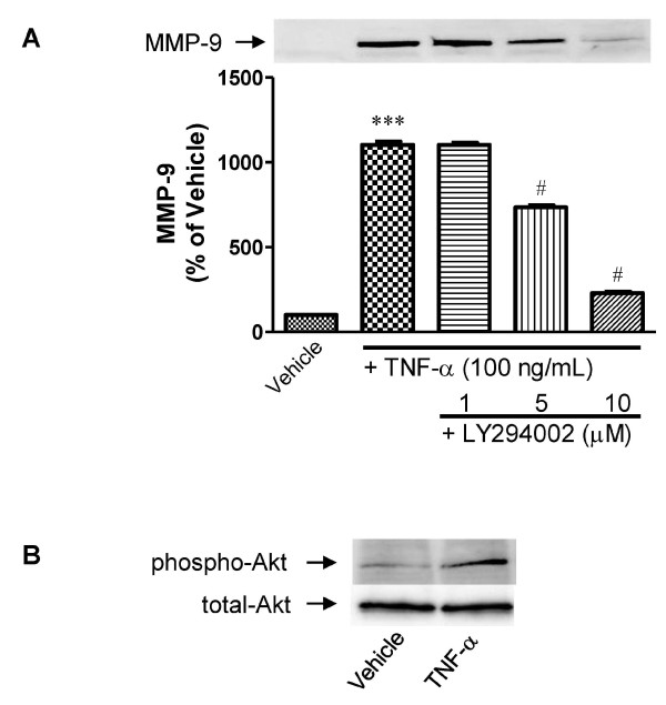
The culture supernatants were collected from primary cultures of rat brain endothelial cells, pericytes, or astrocytes. MMP-9 activities and levels in the supernatants were measured by gelatin zymography and western blot, respectively. The involvement of signaling molecules including mitogen-activated protein kinases (MAPKs) and phosphoinositide-3-kinase (PI3K)/Akt in the mediation of tumor necrosis factor (TNF)-α-induced MMP-9 release was examined using specific inhibitors. The functional activity of MMP-9 was evaluated by a cell migration assay.
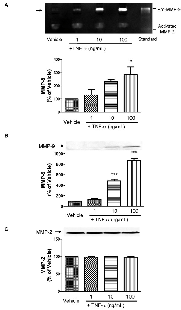
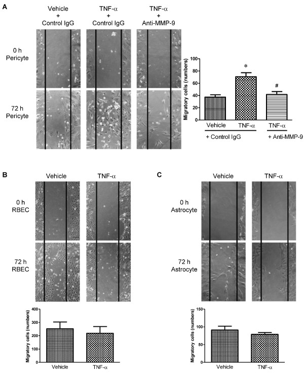
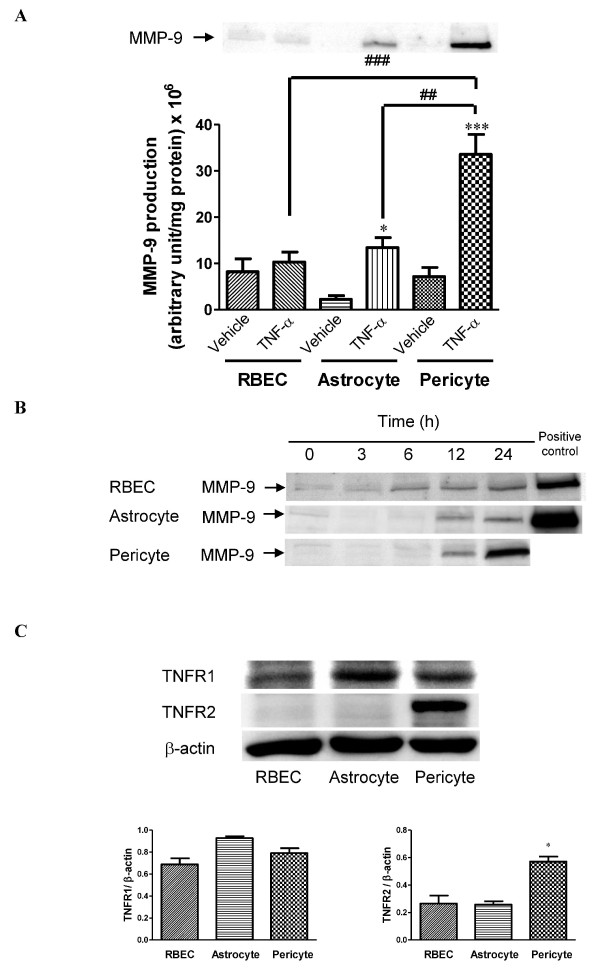
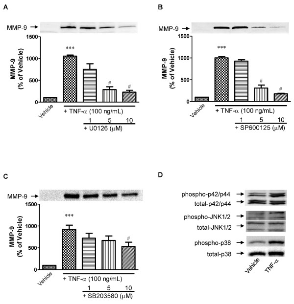
Zymographic and western blot analyses demonstrated that TNF-α stimulated pericytes to release MMP-9, and this release was much higher than from BMECs or astrocytes. Other inflammatory mediators [interleukin (IL)-1β, interferon-γ, IL-6 and lipopolysaccharide] failed to induce MMP-9 release from pericytes. TNF-α-induced MMP-9 release from pericytes was found to be mediated by MAPKs and PI3K. Scratch wound healing assay showed that in contrast to BMECs and astrocytes the extent of pericyte migration was significantly increased by TNF-α. This pericyte migration was inhibited by anti-MMP-9 antibody.
These findings suggest that pericytes are most sensitive to TNF-α in terms of MMP-9 release, and are the major source of MMP-9 at the BBB. This pericyte-derived MMP-9 initiated cellular migration of pericytes, which might be involved in pericyte loss in the damaged BBB.
在神经炎症性疾病中,血浆和大脑中基质金属蛋白酶(MMP)-9 的增加与血脑屏障(BBB)的破坏通过蛋白水解活性有关。MMP-9 存在于脑微血管及其附近,其中脑微血管内皮细胞(BMECs)、周细胞和星形胶质细胞构成了 BBB。关于 MMP-9 在 BBB 中的细胞来源和作用知之甚少。在这里,我们使用从大鼠脑中分离的原代培养物,检查了周细胞在炎症介质作用下释放 MMP-9 和迁移的能力,与 BMECs 和星形胶质细胞进行了比较。
从大鼠脑内皮细胞、周细胞或星形胶质细胞的原代培养物中收集培养上清液。通过明胶酶谱法和 Western blot 分别测量上清液中的 MMP-9 活性和水平。使用特异性抑制剂检查包括丝裂原活化蛋白激酶(MAPKs)和磷酸肌醇 3-激酶(PI3K)/Akt 在内的信号分子在介导肿瘤坏死因子(TNF)-α诱导的 MMP-9 释放中的作用。通过细胞迁移测定评估 MMP-9 的功能活性。
明胶酶谱和 Western blot 分析表明,TNF-α刺激周细胞释放 MMP-9,释放量远高于 BMECs 或星形胶质细胞。其他炎症介质[白细胞介素(IL)-1β、干扰素-γ、IL-6 和脂多糖]未能诱导周细胞释放 MMP-9。发现 TNF-α诱导的周细胞 MMP-9 释放是由 MAPKs 和 PI3K 介导的。划痕愈合试验表明,与 BMECs 和星形胶质细胞相比,TNF-α显著增加了周细胞的迁移程度。这种周细胞迁移被抗 MMP-9 抗体抑制。
这些发现表明,周细胞在 MMP-9 释放方面对 TNF-α最敏感,是 BBB 中 MMP-9 的主要来源。这种周细胞衍生的 MMP-9 引发了周细胞的细胞迁移,这可能与受损 BBB 中周细胞的丧失有关。