Nah Kyung-Soo
Department of Oral and Maxillofacial Radiology, School of Dentistry, Pusan National University, Busan, Korea.
Imaging Sci Dent. 2011 Sep;41(3):107-13. doi: 10.5624/isd.2011.41.3.107. Epub 2011 Sep 15.
The purpose of this study was to present the clinical features of a case series of osteomas in the craniofacial region and to compare them with those described in the dental literatures.
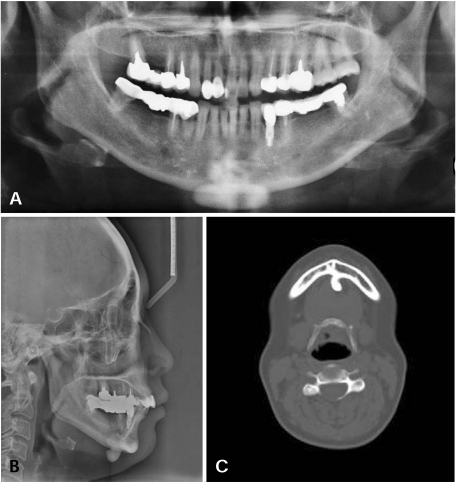
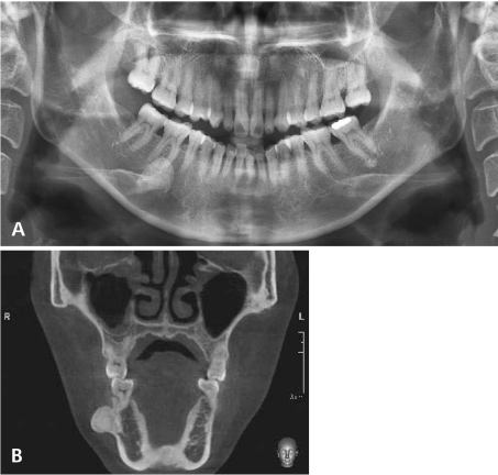
A retrospective study of 18 patients diagnosed with osteomas in the craniofacial region was performed. The age, gender, location, symptoms, and the radiological findings were recorded.
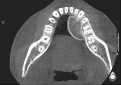
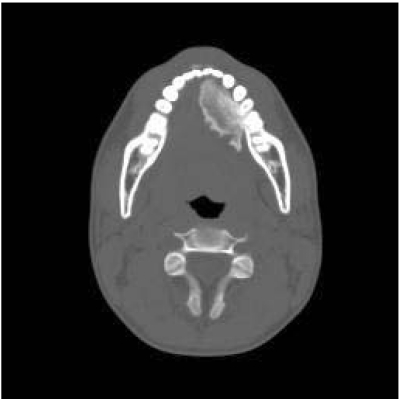
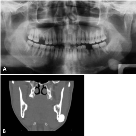
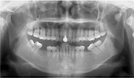
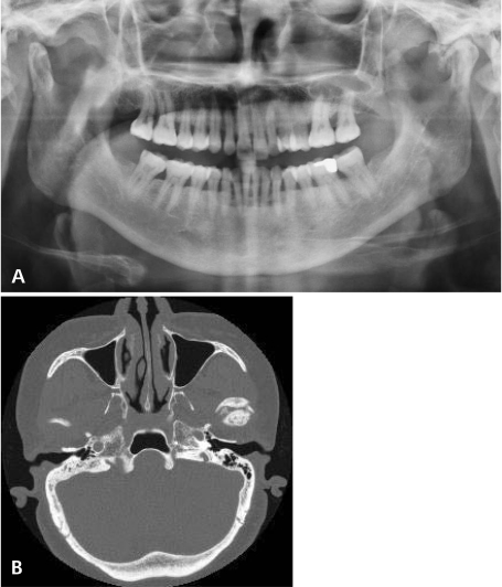
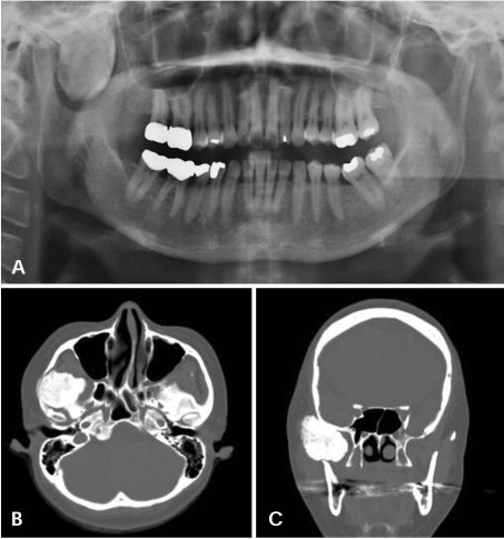
There were 13 women and 5 men from 18 years to 69 years of age (mean age, 42±27 years). Fourteen osteomas were found in the mandible (78%), two in frontal sinus, one in sphenoid bone, and one in maxilla.
Osteomas are benign tumors composed of mature compact bone or cancellous bone. They are essentially restricted to the craniofacial skeleton and rarely, if ever, are diagnosed in other bones.
本研究的目的是呈现一组颅面部骨瘤病例的临床特征,并将其与牙科文献中描述的特征进行比较。
对18例诊断为颅面部骨瘤的患者进行回顾性研究。记录患者的年龄、性别、部位、症状及影像学表现。
18例患者中,女性13例,男性5例,年龄18岁至69岁(平均年龄42±27岁)。14例骨瘤位于下颌骨(78%),2例位于额窦,1例位于蝶骨,1例位于上颌骨。
骨瘤是由成熟的密质骨或松质骨组成的良性肿瘤。它们基本上局限于颅面骨骼,极少(如果有的话)在其他骨骼中被诊断出来。