Paracelsus Medical University, Salzburg, Austria.
Arthritis Care Res (Hoboken). 2012 Nov;64(11):1681-90. doi: 10.1002/acr.21719.
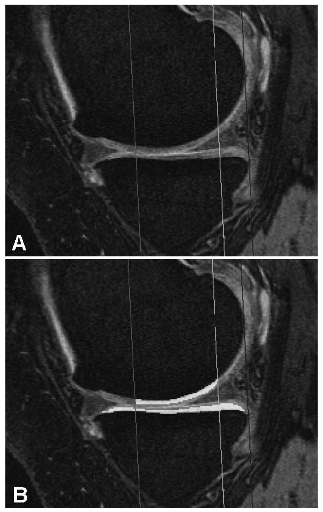
To determine whether the presence of definite osteophytes (in the absence of joint space narrowing [JSN]) on radiographs is associated with (subregional) increases in cartilage thickness in a within-person, between-knee cross-sectional comparison of participants in the Osteoarthritis Initiative. Based on previous results, the external weight-bearing medial femoral condyle (ecMF) and external weight-bearing lateral femoral condyle (ecLF) subregions were selected as primary end points.
Both knees of 61 Osteoarthritis Initiative participants (n = 4,796) displayed definite tibial or femoral marginal osteophytes and no JSN in 1 knee, and no signs of radiographic osteoarthritis (OA) in the contralateral knee; this was confirmed by an expert central reader. In these participants, cartilage thickness was measured in 16 femorotibial subregions of each knee, based on sagittal double-echo steady-state with water excitation magnetic resonance images. Location-specific joint space width from fixed-flexion radiographs was determined using dedicated software. Location-specific associations of osteophytes with cartilage thickness were evaluated using paired t-tests and mixed-effects models.
Of the 61 participants, 48% had only medial osteophytes, 36% only lateral osteophytes, and 16% bicompartmental osteophytes. The knees with osteophytes had significantly thicker cartilage than contralateral knees without osteophytes in the ecMF (mean ± SD +71 ± 223 μmoles, equivalent to an increase of +5.5%; P = 0.015) and ecLF (mean ± SD +64 ± 195 μmoles, +4.1%; P = 0.013). No significant differences between knees were noted in other subregions or in joint space width. Cartilage thickness in the ecMF and ecLF was significantly associated with tibial osteophytes in the same (medial or lateral) compartment (P = 0.003).
The knees with early radiographic OA display thicker cartilage than (contralateral) knees without radiographic findings of OA, specifically in the external femoral subregions of compartments with marginal osteophytes.
在骨关节炎倡议(Osteoarthritis Initiative)参与者的个体内、双膝横断面比较中,确定放射照片上是否存在明确的骨赘(无关节间隙变窄[JSN])是否与软骨厚度的(亚区)增加相关。基于先前的结果,选择外部负重股骨内侧髁(ecMF)和外部负重股骨外侧髁(ecLF)亚区作为主要终点。
61 名骨关节炎倡议参与者(n=4796)的双侧膝关节中,一侧膝关节存在明确的胫骨或股骨边缘骨赘且无 JSN,而对侧膝关节无放射学骨关节炎(OA)迹象;这是由一位专家中心读者确认的。在这些参与者中,根据矢状面双回波稳态水激发磁共振成像,测量了每侧膝关节 16 个股骨胫骨亚区的软骨厚度。使用专用软件从固定弯曲射线照片确定特定部位的关节间隙宽度。使用配对 t 检验和混合效应模型评估骨赘与软骨厚度的特定部位关联。
在 61 名参与者中,48%仅存在内侧骨赘,36%仅存在外侧骨赘,16%存在双间室骨赘。存在骨赘的膝关节的软骨厚度明显比没有骨赘的对侧膝关节厚,在 ecMF(平均±SD+71±223μmoles,相当于增加 5.5%;P=0.015)和 ecLF(平均±SD+64±195μmoles,增加 4.1%;P=0.013)。在其他亚区或关节间隙宽度方面,膝关节之间没有显著差异。ecMF 和 ecLF 中的软骨厚度与同一(内侧或外侧)间室的胫骨骨赘显著相关(P=0.003)。
有早期放射学 OA 的膝关节的软骨厚度比(对侧)没有放射学 OA 发现的膝关节厚,特别是在有边缘骨赘的间室的外部股骨亚区。