Section of Human Physiology, University of Perugia School of Medicine, Perugia, Italy.
Neurobiol Dis. 2012 Sep;47(3):310-21. doi: 10.1016/j.nbd.2012.05.002. Epub 2012 May 17.
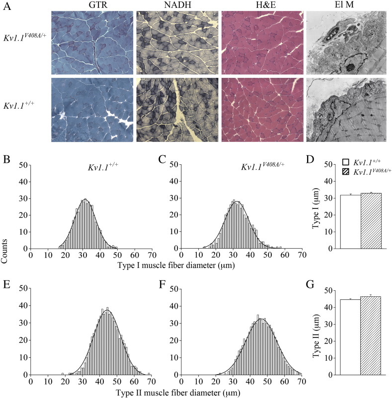
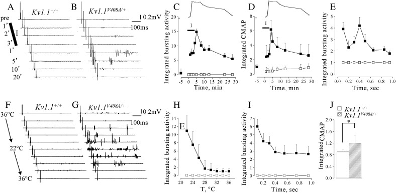
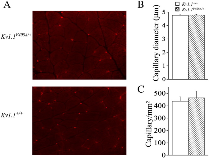
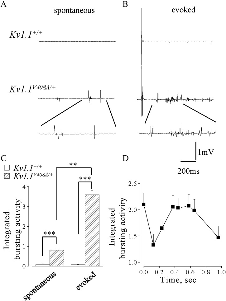
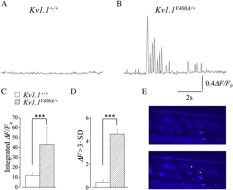
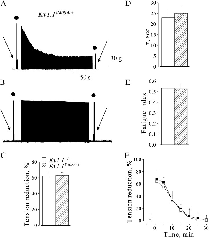
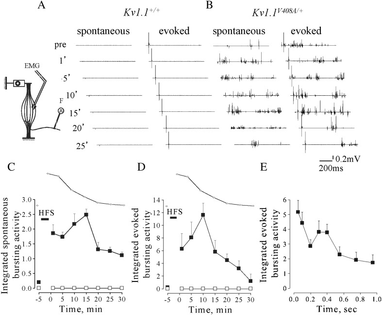
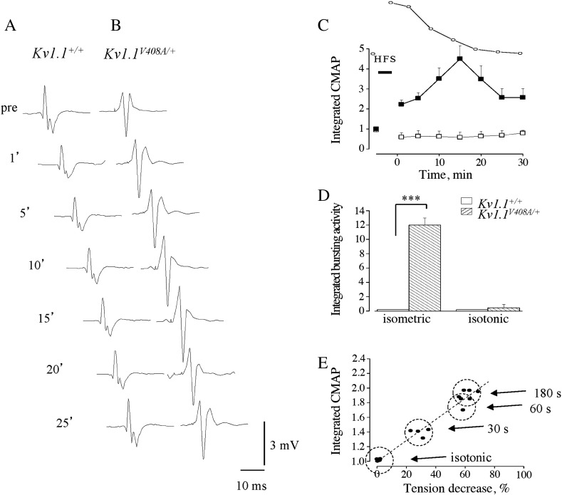
Episodic ataxia type 1 (EA1) is an autosomal dominant neurological disorder characterized by myokymia and attacks of ataxic gait often precipitated by stress. Several genetic mutations have been identified in the Shaker-like K(+) channel Kv1.1 (KCNA1) of EA1 individuals, including V408A, which result in remarkable channel dysfunction. By inserting the heterozygous V408A, mutation in one Kv1.1 allele, a mouse model of EA1 has been generated (Kv1.1(V408A/+)). Here, we investigated the neuromuscular transmission of Kv1.1(V408A/+) ataxic mice and their susceptibility to physiologically relevant stressors. By using in vivo preparations of lateral gastrocnemius (LG) nerve-muscle from Kv1.1(+/+) and Kv1.1(V408A/+) mice, we show that the mutant animals exhibit spontaneous myokymic discharges consisting of repeated singlets, duplets or multiplets, despite motor nerve axotomy. Two-photon laser scanning microscopy from the motor nerve, ex vivo, revealed spontaneous Ca(2+) signals that occurred abnormally only in preparations dissected from Kv1.1(V408A/+) mice. Spontaneous bursting activity, as well as that evoked by sciatic nerve stimulation, was exacerbated by muscle fatigue, ischemia and low temperatures. These stressors also increased the amplitude of compound muscle action potential. Such abnormal neuromuscular transmission did not alter fiber type composition, neuromuscular junction and vascularization of LG muscle, analyzed by light and electron microscopy. Taken together these findings provide direct evidence that identifies the motor nerve as an important generator of myokymic activity, that dysfunction of Kv1.1 channels alters Ca(2+) homeostasis in motor axons, and also strongly suggest that muscle fatigue contributes more than PNS fatigue to exacerbate the myokymia/neuromyotonia phenotype. More broadly, this study points out that juxtaparanodal K(+) channels composed of Kv1.1 subunits exert an important role in dampening the excitability of motor nerve axons during fatigue or ischemic insult.
发作性共济失调 1 型(EA1)是一种常染色体显性遗传性神经系统疾病,其特征为肌纤维震颤和共济失调步态发作,常由应激引发。在 EA1 个体的 Shaker 样 K(+)通道 Kv1.1(KCNA1)中已鉴定出几种基因突变,包括 V408A,其导致显著的通道功能障碍。通过插入杂合 V408A,即在一个 Kv1.1 等位基因中突变,已经生成了 EA1 的小鼠模型(Kv1.1(V408A/+))。在此,我们研究了 Kv1.1(V408A/+)共济失调小鼠的神经肌肉传递及其对生理相关应激源的易感性。通过使用 Kv1.1(+/+)和 Kv1.1(V408A/+)小鼠的外侧腓肠肌(LG)神经-肌肉的体内标本,我们显示尽管运动神经轴突切断,突变动物仍表现出由重复单、双或多肌纤维颤搐组成的自发性肌纤维震颤放电。离体的双光子激光扫描显微镜从运动神经中揭示,仅在从 Kv1.1(V408A/+)小鼠分离的标本中才会异常发生自发性 Ca(2+)信号。自发性爆发活动以及由坐骨神经刺激引起的活动,在肌肉疲劳、缺血和低温下加剧。这些应激源还增加了复合肌肉动作电位的幅度。通过光和电子显微镜分析,这种异常的神经肌肉传递并没有改变 LG 肌肉的纤维类型组成、神经肌肉接头和血管化。总之,这些发现提供了直接证据,确定运动神经是肌纤维震颤活动的重要发生源,Kv1.1 通道的功能障碍改变了运动轴突中的 Ca(2+)稳态,并且强烈表明肌肉疲劳比周围神经疲劳更能加剧肌纤维震颤/神经肌强直表型。更广泛地说,这项研究指出,由 Kv1.1 亚基组成的近旁节段 K(+)通道在疲劳或缺血损伤期间对降低运动神经轴突的兴奋性具有重要作用。