Department of Emergency, Third Hospital of Hebei Medical University, Shijiazhuang 050051, China.
Int J Med Sci. 2012;9(6):413-23. doi: 10.7150/ijms.4514. Epub 2012 Jul 21.
To determine the role of microRNA 21(miR-21) on left ventricular remodeling of rat heart with ischemia-reperfusion (I/R) injury and to investigate the underlying mechanism of miR-21 mediated myocardium protection.
Rats were randomly divided into three groups: an I/R model group with Ad-GFP (Ad-GFP group), an I/R model group with Ad-miR-21 (Ad-miR-21 group) and a sham-surgery group. Changes in hemodynamic parameters were recorded at 1 week after I/R. Histological diagnosis was achieved by hematoxylin and eosin (H&E). Left ventricular (LV) dimensions, myocardial infarct size, LV/BW, collagen type Ⅰ, type Ⅲ and PCNA positive cells were measured. Primary cultures of neonatal rat cardiac ventricular myocytes were performed and cell ischemic injury was induced by hypoxia in a serum- and glucose-free medium, and reoxygenation (H/R). MiR-21 inhibitor and pre-miR-21 were respectively added to the culture medium for the miR-21 knockdown and for the miR-21 up-regulation. qRT-PCR was used to determine the miR-21 levels in cultured cells. Flow cytometry was performed to examine the cell apoptosis.
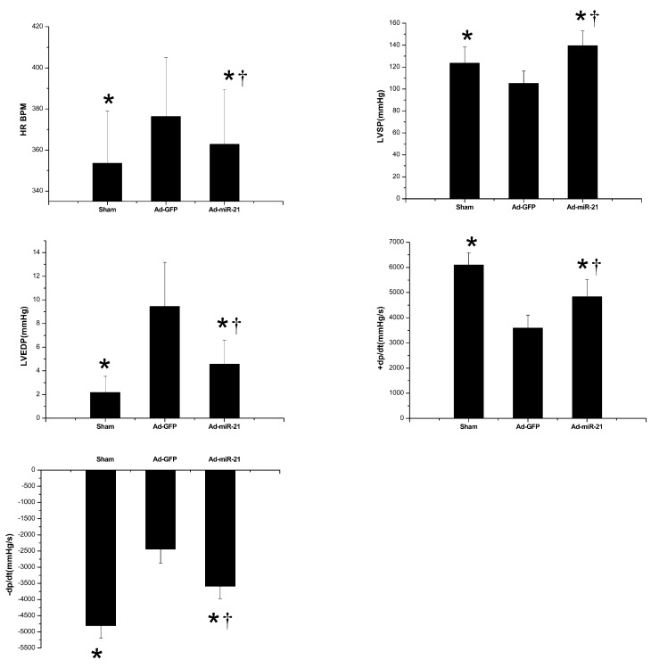
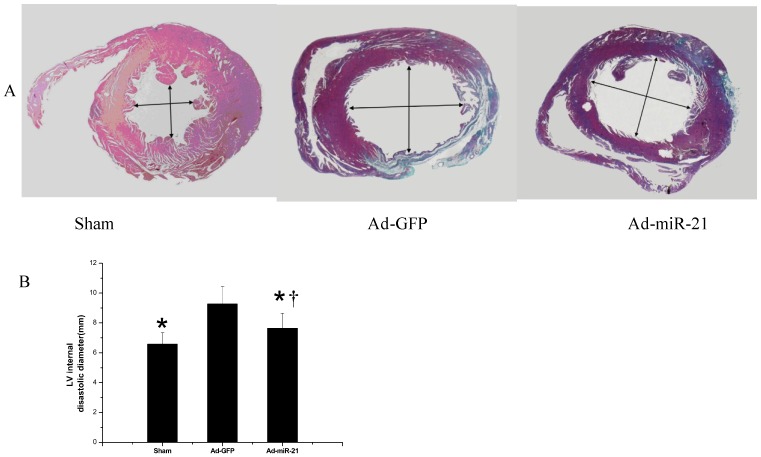
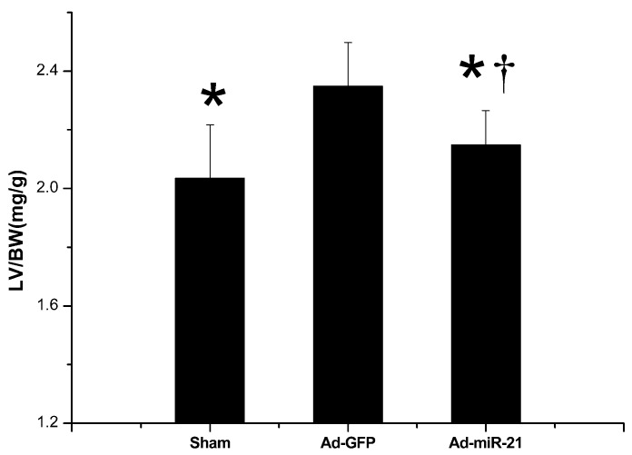
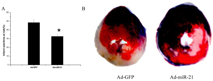
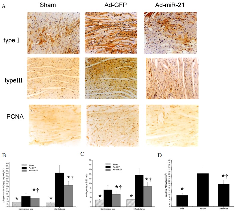
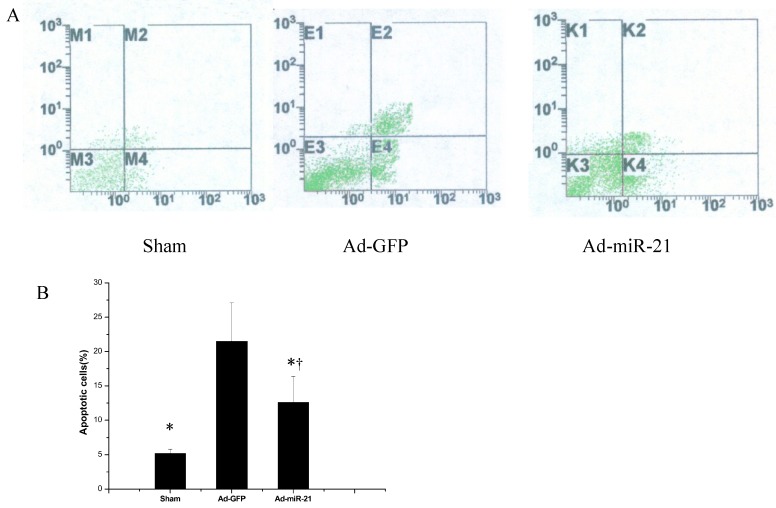
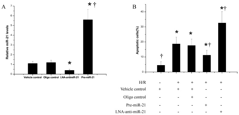
In the Ad-miR-21 group, LV dimensions, myocardial infarct size, LV/BW, collagen type Ⅰ, type Ⅲ and PCNA positive cells all significantly decreased compared with the Ad-GFP group. At 1 week after I/R, the Ad-miR-21 significantly improved LVSP, LV +dp/dt(max), LV - dp/dt(min), and decreased heart rate (HR) and LVEDP compared with the Ad-GFP group. Compared with the Ad-GFP, the cell apoptotic rate significantly decreased in the Ad-miR-21 group. The miR-21 inhibitor exacerbated cardiac myocyte apoptosis and the pre-miR-21 decreased hypoxia/reoxygenation- induced cardiac myocyte apoptosis.
Ad-miR-21 improves LV remodeling and decreases the apoptosis of myocardial cells, suggesting the possible mechanism by which Ad-miR-21 functions in protecting against I/R injury.
确定微小 RNA 21(miR-21)在缺血再灌注(I/R)损伤大鼠心脏左心室重构中的作用,并探讨 miR-21 介导心肌保护的潜在机制。
将大鼠随机分为三组:携带 Ad-GFP(Ad-GFP 组)的 I/R 模型组、携带 Ad-miR-21(Ad-miR-21 组)的 I/R 模型组和假手术组。I/R 后 1 周记录血流动力学参数变化。苏木精-伊红(H&E)染色进行组织学诊断。测量左心室(LV)尺寸、心肌梗死面积、LV/BW、胶原 I、III 型和 PCNA 阳性细胞。原代培养新生大鼠心室肌细胞,在无血清和低糖培养基中缺氧诱导细胞缺血损伤,再复氧(H/R)。分别向培养基中加入 miR-21 抑制剂和 pre-miR-21 以实现 miR-21 的敲低和上调。qRT-PCR 用于测定培养细胞中的 miR-21 水平。流式细胞术检测细胞凋亡。
在 Ad-miR-21 组,与 Ad-GFP 组相比,LV 尺寸、心肌梗死面积、LV/BW、胶原 I、III 型和 PCNA 阳性细胞均显著减少。I/R 后 1 周,与 Ad-GFP 组相比,Ad-miR-21 显著提高了 LVSP、LV+dp/dt(max)、LV-dp/dt(min),并降低了心率(HR)和 LVEDP。与 Ad-GFP 相比,Ad-miR-21 组细胞凋亡率显著降低。miR-21 抑制剂加重了心肌细胞凋亡,而 pre-miR-21 减少了缺氧/复氧诱导的心肌细胞凋亡。
Ad-miR-21 改善 LV 重构并减少心肌细胞凋亡,提示 Ad-miR-21 发挥抗 I/R 损伤作用的可能机制。